
Science Lab
Science Lab
Das Wissensportal von Leica Microsystems bietet Ihnen Wissens- und Lehrmaterial zu den Themen der Mikroskopie. Die Inhalte sind so konzipiert, dass sie Einsteiger, erfahrene Praktiker und Wissenschaftler gleichermaßen bei ihrem alltäglichen Vorgehen und Experimenten unterstützen. Entdecken Sie interaktive Tutorials und Anwendungsberichte, erfahren Sie mehr über die Grundlagen der Mikroskopie und High-End-Technologien - werden Sie Teil der Science Lab Community und teilen Sie Ihr Wissen!
Loading...
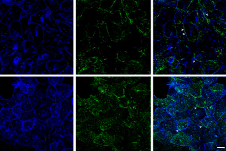
Improvement of Imaging Techniques to Understand Organelle Membrane Cell Dynamics
Understanding cell functions in normal and tumorous tissue is a key factor in advancing research of potential treatment strategies and understanding why some treatments might fail. Single-cell…
Loading...

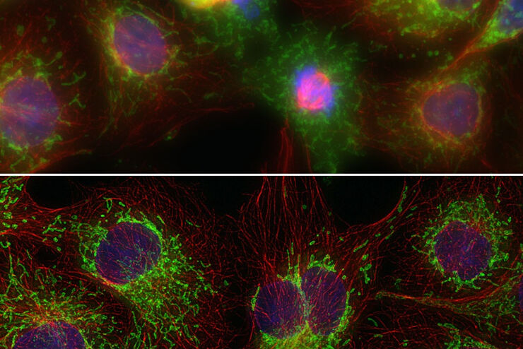
Image Gallery: THUNDER Imager
To help you answer important scientific questions, THUNDER Imagers eliminate the out-of-focus blur that clouds the view of thick samples when using camera-based fluorescence microscopes. They achieve…
Loading...

Studying Human Brain Development and Disease
Neural spheroids created from human induced pluripotent stem cells (iPSCs) provide effective and novel tools for studying brain development, as well as the underlying pathological mechanisms of…
Loading...

An Introduction to Computational Clearing
Many software packages include background subtraction algorithms to enhance the contrast of features in the image by reducing background noise. The most common methods used to remove background noise…
Loading...
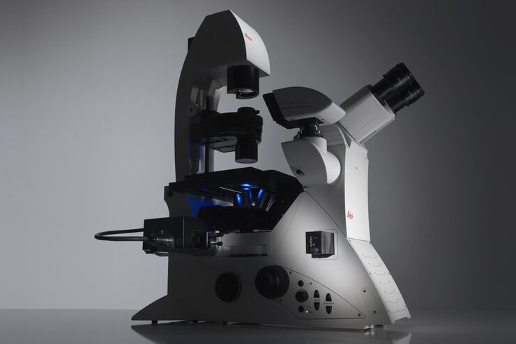
Factors to Consider When Selecting a Research Microscope
An optical microscope is often one of the central devices in a life-science research lab. It can be used for various applications which shed light on many scientific questions. Thereby the…
Loading...
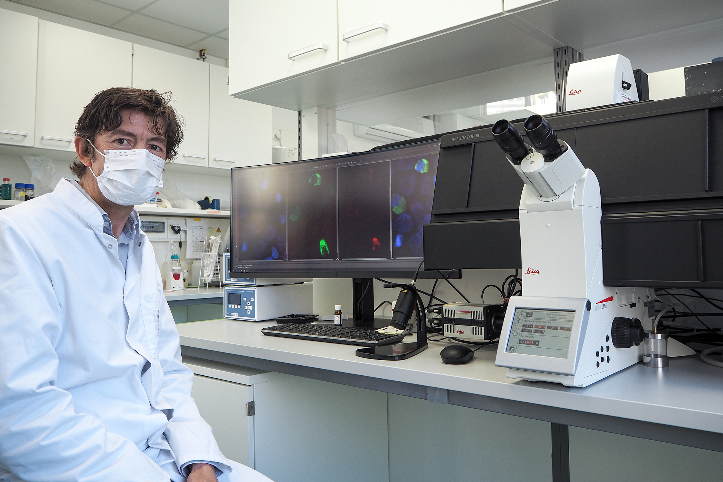
Microscopy in Virology
The coronavirus SARS-CoV-2, causing the Covid-19 disease effects our world in all aspects. Research to find immunization and treatment methods, in other words to fight this virus, gained highest…
Loading...
Computational Clearing - Enhance 3D Specimen Imaging
This webinar is designed to clarify crucial specifications that contribute to THUNDER Imagers' transformative visualization of 3D samples and improvements within a researcher's imaging-related…
Loading...
THUNDER Imagers: High Performance, Versatility and Ease-of-Use for your Everyday Imaging Workflows
This webinar will showcase the versatility and performance of THUNDER Imagers in many different life science applications: from counting nuclei in retina sections and RNA molecules in cancer tissue…
Loading...
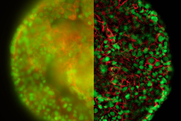
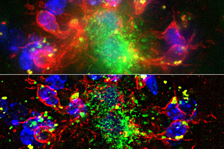
Real Time Images of 3D Specimens with Sharp Contrast Free of Haze
THUNDER Imagers deliver in real time images of 3D specimens with sharp contrast, free of the haze or out-of-focus blur typical of widefield systems. They can even image clearly places deep inside a…