
Science Lab
Science Lab
Das Wissensportal von Leica Microsystems bietet Ihnen Wissens- und Lehrmaterial zu den Themen der Mikroskopie. Die Inhalte sind so konzipiert, dass sie Einsteiger, erfahrene Praktiker und Wissenschaftler gleichermaßen bei ihrem alltäglichen Vorgehen und Experimenten unterstützen. Entdecken Sie interaktive Tutorials und Anwendungsberichte, erfahren Sie mehr über die Grundlagen der Mikroskopie und High-End-Technologien - werden Sie Teil der Science Lab Community und teilen Sie Ihr Wissen!
Loading...

How AR Helps in the Surgical Treatment of Moyamoya Disease
Moyamoya disease is a rare chronic occlusive cerebrovascular disorder characterized by progressive stenosis in the terminal portion of the internal carotid artery and an abnormal vascular network at…
Loading...

Using GLOW800 AR in Radial Forearm Flap Free Phalloplasty
In this video, Chief Microsurgeon Professor Küntscher and his team perform a radial forearm free flap phalloplasty and use ICG fluorescence imaging to show the blood flow in the whole flap from the…
Loading...
 expressed in the sensory neurons.")
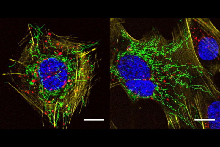
Fast, High-contrast 3D Imaging of Sensory Neurons
This article discusses how fast, high-contrast 3D imaging of dorsal root ganglion (DRG) tissue with a THUNDER Imager Tissue using large volume computational clearing (LVCC) allows sensory neurons to…
Loading...
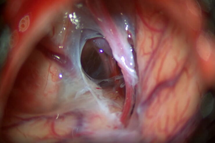
 or Minor’s syndrome")
Operative Intervention bei Bogengangsdehiszenz
Minor’s disease, also called Superior Semicircular Canal Dehiscence (SSCD) or Minor’s syndrome, is a rare disorder of the inner ear that affects hearing and balance. The disease is characterized by…
Loading...
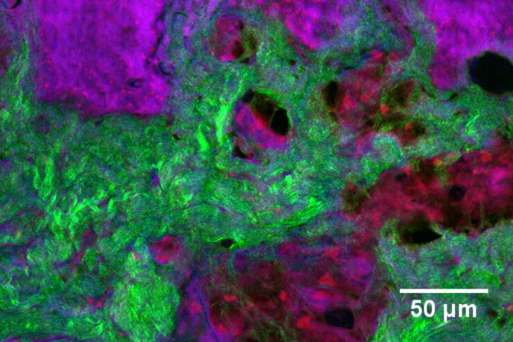
A Versatile Palette of Fluorescent Probes
Researchers at the Max Planck Institute for Medical Research in Heidelberg have developed a general strategy to synthesize live-cell compatible fluorogenic probes, and the result are the new MaP (Max…
Loading...
Formulated Product Characterization with SRS Microscopy
Creams, pastes, gels, emulsions, and tablets are ubiquitous across a wide range of manufacturing sectors from pharmaceuticals and consumer health products to agrochemicals and paint. To improve…
Loading...
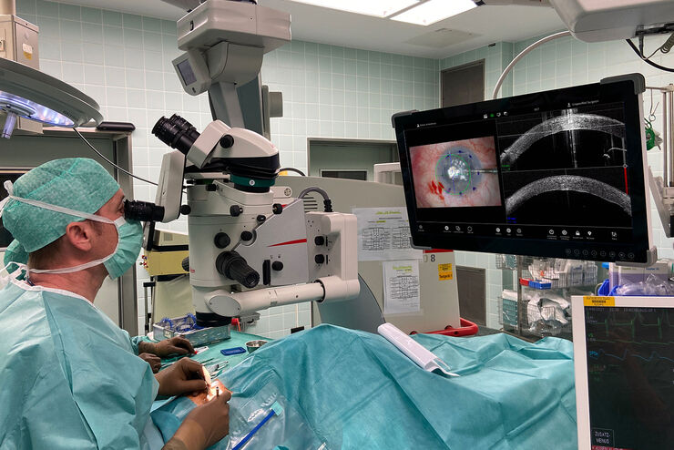
Proveo 8 with intraoperative OCT – a User Evaluation in an University Setting
Optical coherence tomography (OCT) makes structures in the eye visible that lie beneath the surface. When OCT is used intraoperatively, surgeons gain insight into possible pathological changes in the…
Loading...
Optimal Visualization in Brain Surgery
This case study “Treatment of the Galassi type III arachnoid cyst with the M530 OHX surgical microscope from Leica Microsystems” documents the procedure step by step and shows the visualization…
Loading...
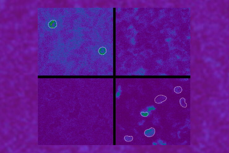
Spectroscopic Evaluation of Red Blood Cells
Hemoglobinopathies are a major healthcare problem. This study presents a possible diagnostic tool for thalassemia which is based on confocal spectroscopy. This approach exploits spectral detection and…