
Science Lab
Science Lab
Das Wissensportal von Leica Microsystems bietet Ihnen Wissens- und Lehrmaterial zu den Themen der Mikroskopie. Die Inhalte sind so konzipiert, dass sie Einsteiger, erfahrene Praktiker und Wissenschaftler gleichermaßen bei ihrem alltäglichen Vorgehen und Experimenten unterstützen. Entdecken Sie interaktive Tutorials und Anwendungsberichte, erfahren Sie mehr über die Grundlagen der Mikroskopie und High-End-Technologien - werden Sie Teil der Science Lab Community und teilen Sie Ihr Wissen!
Loading...

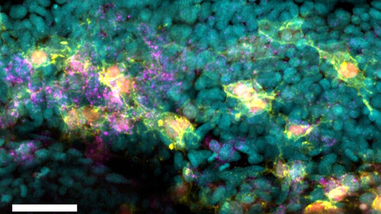
Unlocking Insights in Complex and Dense Neuron Images Guided by AI
The latest advancement in Aivia AI image analysis software provides improved soma detection, additional flexibility in neuron tracing, 3D relational measurement including Sholl analysis and more.
Loading...
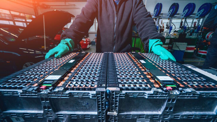
How to Prepare and Analyse Battery Samples with Electron Microscopy
This workshop covers the sample preparation process for lithium and novel battery sample analysis, as well as other semiconductor samples requiring high-resolution cross-section imaging.
Loading...

Die Rolle des Eisenstoffwechsels bei der Krebsentwicklung
Der Eisenstoffwechsel spielt eine Rolle bei der Entstehung und dem Fortschreiten von Krebs und beeinflusst die Immunreaktion. Zu verstehen, wie Eisen Krebs und das Immunsystem beeinflusst, kann die…
Loading...

AR Fluorescence in Aneurysm Clipping and AVM Surgery
Discover how GLOW800 Augmented Reality fluorescence supports neurovascular surgical procedures and in particular aneurysm clipping and AVM surgery.
Loading...

How to Image Histological and Fluorescent Samples with One System
VIDEO ON DEMAND - How to image histological and fluorescent samples with one system. FluoSync, the new technology embedded into Mica enables the imaging of both histological staining and fluorescence…
Loading...

Surgical Management of High-Grade Gliomas
Learn about the surgical management of high-grade gliomas and how to expand the extent of resection intra-operatively using tools such as 5-ALA fluorescence.
Loading...
How to Radically Simplify Workflows in Your Imaging Facility
VIDEO ON DEMAND - How to radically simplify imaging workflows and generate meaningful results with less time and effort using a highly automated microscope that unites widefield and confocal imaging.
Loading...

Advancing Surgery in Pediatric Brain Tumors
Learn how fluorescence-guided surgery supports pediatric brain tumor resection and improves precision through literature review and clinical cases.
Loading...

Five-color FLIM-STED with One Depletion Laser
Webinar on five-color STED with a single depletion laser and fluorescence lifetime phasor separation.