
Science Lab
Science Lab
Bienvenue sur le portail de connaissances de Leica Microsystems. Vous y trouverez des recherches scientifiques et du matériel didactique sur le thème de la microscopie. Le portail aide les débutants, les praticiens expérimentés et les scientifiques dans leur travail quotidien et leurs expériences. Explorez les didacticiels interactifs et les notes d'application, découvrez les bases de la microscopie ainsi que les technologies de pointe. Faites partie de la communauté Science Lab et partagez votre expertise.
Filter articles
Tags
Type de publication
Produits
Loading...

Organismes Modèles dans la Recherche
Un organisme modèle désigne une espèce utilisée par les chercheurs pour étudier des processus biologiques spécifiques. Ils présentent des caractéristiques génétiques similaires à celles de l’homme et…
Loading...

Recherche sur le cancer
Le cancer est une maladie complexe et hétérogène causée par des cellules déficientes dans la régulation de la croissance. Les modifications génétiques et épigénétiques d’une cellule ou d’un groupe de…
Loading...

Introduction to Widefield Microscopy
This article gives an introduction to widefield microscopy, one of the most basic and commonly used microscopy techniques. It also shows the basic differences between widefield and confocal…
Loading...
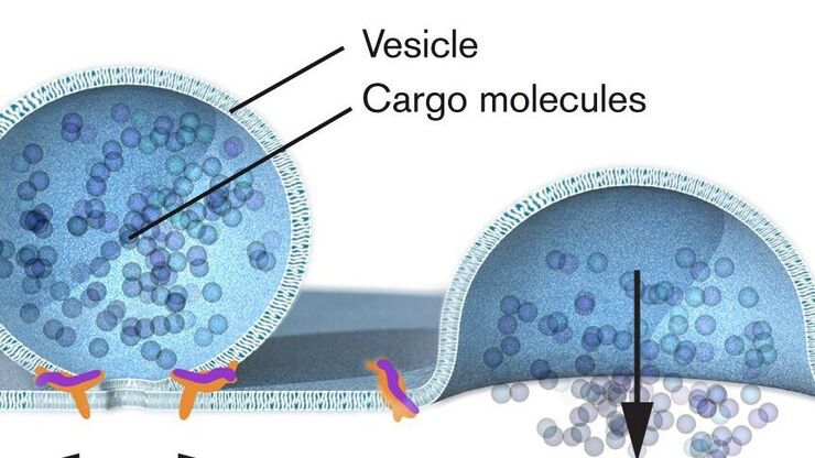
Nobel Prize 2013 in Physiology or Medicine for Discoveries of the Machinery Regulating Vesicle Traffic
On October 7th 2013, The Nobel Assembly at Karolinska Institutet has decided to award The Nobel Prize in Physiology or Medicine 2012 jointly to James E. Rothman, Randy W. Schekman and Thomas C. Südhof…
Loading...

Nobel Prize 2012 in Physiology or Medicine for Stem Cell Research
The Nobel Prize recognizes two scientists who discovered that mature, specialised cells can be reprogrammed to become immature cells capable of developing into all tissues of the body. Their findings…
Loading...
 inoculated with cowpea mosaic virus (CPMV) containing the GFP-gene inserted between the movement protein (MP) and the capsid proteins (CPs) in the viral RNA 2")
Introduction to Live-Cell Imaging
The understanding of complex and fast cellular dynamics is an important step to get insight into biological processes. Therefore, today’s life science research more and more demands studying…
