
Industrial Microscopy
Industrial Microscopy
Dive deep into detailed articles and webinars focusing on efficient inspection, optimized workflows, and ergonomic comfort in industrial and pathological contexts. Topics covered include quality control, materials analysis, microscopy in pathology, among many others. This is the place where you get valuable insights into using cutting-edge technologies for improving precision and efficiency in manufacturing processes as well as accurate pathological diagnosis and research.
Digital Microscopy in Forensics
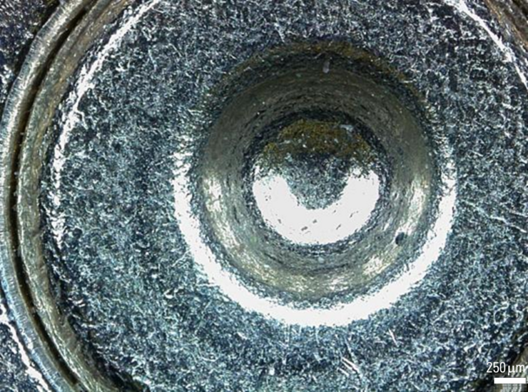
Forensic experts work with a broad range of microscopes to examine evidence from firearms and tool marks, documents, forensic or legal medicine, hair and fibers as well as glass and paint. Digital…
Life Science Imaging with DVM6 Digital Microscope
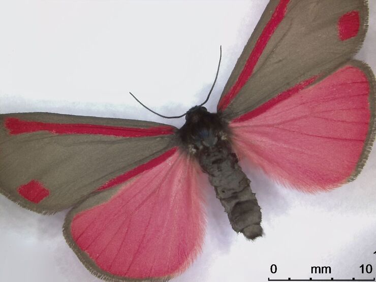
Digital microscopes can be a great help in life science applications such as the documentation in botany, entomology studies and crop science, or the digitization of museum collections. The image…

What You Always Wanted to Know About Digital Microscopy, but Never Got Around to Asking
Digital microscopy is one of the buzz words in microscopy – and there are a couple of facts that are useful to know. Georg Schlaffer, Product Manager with Leica Microsystems, has often been asked…

Immersion Freezing for Cryo-Transmission Electron Microscopy: Applications
A well established usage case for cryo-TEM is three-dimensional reconstruction of isolated macromolecules, virus particles, or filaments. These approaches are based on averaging of repetitive…
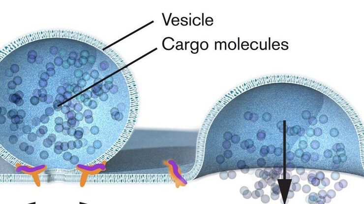
Nobel Prize 2013 in Physiology or Medicine for Discoveries of the Machinery Regulating Vesicle Traffic
On October 7th 2013, The Nobel Assembly at Karolinska Institutet has decided to award The Nobel Prize in Physiology or Medicine 2012 jointly to James E. Rothman, Randy W. Schekman and Thomas C. Südhof…

Nobel Prize 2012 in Physiology or Medicine for Stem Cell Research
The Nobel Prize recognizes two scientists who discovered that mature, specialised cells can be reprogrammed to become immature cells capable of developing into all tissues of the body. Their findings…
 inoculated with cowpea mosaic virus (CPMV) containing the GFP-gene inserted between the movement protein (MP) and the capsid proteins (CPs) in the viral RNA 2")
Introduction to Live-Cell Imaging
The understanding of complex and fast cellular dynamics is an important step to get insight into biological processes. Therefore, today’s life science research more and more demands studying…

Is that Document Genuine or Fake? How do They Identify Fake Documents?
This article shows how forensic experts use microscopy for analysis to identify counterfeit, fake documents, such as ID cards, passports, visas, certificates, etc. Then they know if it is genuine or…
