
Industrial Microscopy
Industrial Microscopy
Dive deep into detailed articles and webinars focusing on efficient inspection, optimized workflows, and ergonomic comfort in industrial and pathological contexts. Topics covered include quality control, materials analysis, microscopy in pathology, among many others. This is the place where you get valuable insights into using cutting-edge technologies for improving precision and efficiency in manufacturing processes as well as accurate pathological diagnosis and research.
Filter articles
Tags
Loading...
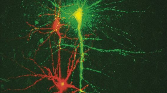
New Standard in Electrophysiology and Deep Tissue Imaging
The function of nerve and muscle cells relies on ionic currents flowing through ion channels. These ion channels play a major role in cell physiology. One way to investigate ion channels is to use…
Loading...

The History of Stereo Microscopy
This article gives an overview on the history of stereo microscopes. The development and evolution from handcrafted instruments (late 16th to mid-18th century) to mass produced ones the last 150…
