
Medical Specialties
Medical Specialties
Explore a comprehensive collection of scientific and clinical resources tailored for HCPs, including peer insights, clinical case studies, and symposia. Designed for neurosurgeons, ophthalmologists, and specialists in Plastic and Reconstructive surgery, ENT, and dentistry. This collection highlights the latest advancements in surgical microscopy. Discover how cutting-edge surgical technologies, such as AR fluorescence, 3D visualization, and intraoperative OCT imaging, empower confident decision-making and precision in complex surgeries.
Brief Introduction to Specimen Trimming
Before ultrathin sectioning a sample with an ultramicrotome it has to be pre-prepared. For this pre-preparation, special attention must be paid to the sample size (size of the section), location of…
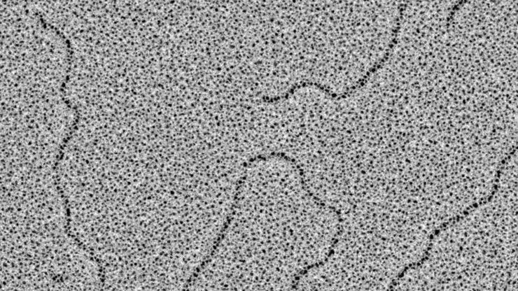
Glycerol Spraying/Platinum Low Angle Rotary Shadowing of DNA with the Leica EM ACE600 e-beam
Glycerol spraying/low angle rotary shadowing (Aebi and Baschong, 2006) is a preparation technique used in biology to visualize structures yielding insufficent contrast with other techniques, due to…

Brief Introduction to Freeze Substitution
Freeze-substitution is a process of dehydration, performed at temperatures low enough to avoid the formation of ice crystals and to circumvent the damaging effects observed after ambient-temperature…

Acousto Optics in True Confocal Spectral Microscope Systems
Acousto-optical elements have successfully replaced planar filters in many positions. The white confocal, regarded as the fully spectrally tunable confocal microscope, was not possible without this…
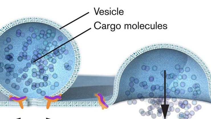
Nobel Prize 2013 in Physiology or Medicine for Discoveries of the Machinery Regulating Vesicle Traffic
On October 7th 2013, The Nobel Assembly at Karolinska Institutet has decided to award The Nobel Prize in Physiology or Medicine 2012 jointly to James E. Rothman, Randy W. Schekman and Thomas C. Südhof…
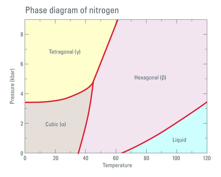
Thermodynamic Considerations Regarding the LN2 in a High Pressure Freezer
Employing liquid nitrogen (LN2) as a coolant in the complex process of high pressure freezing raises certain considerations regarding phase transition not only of the liquid sample to be frozen but…

Brief Introduction to Contrasting for EM Sample Preparation
Since the contrast in the electron microscope depends primarily on the differences in the electron density of the organic molecules in the cell, the efficiency of a stain is determined by the atomic…

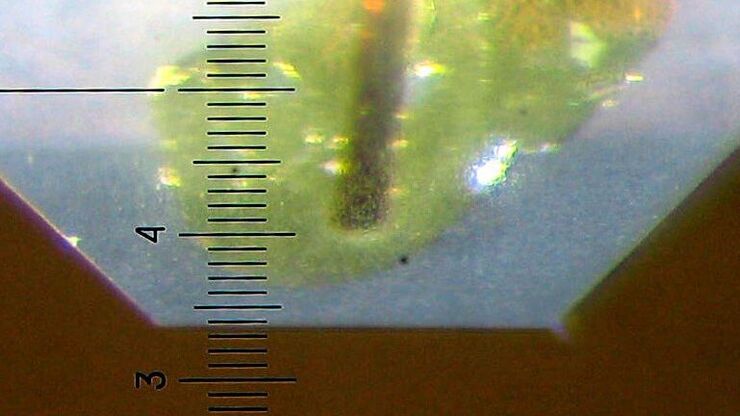
Brief Introduction to Glass Knifemaking for Electron and Light Microscope Applications
Glass knives are used in an ultramicrotome to cut ultrathin slices of samples for electron and light microscope applications. For resin and for cryo sections (Tokuyasu samples) the knife edge must be…

Brief Introduction to Coating Technology for Electron Microscopy
Coating of samples is required in the field of electron microscopy to enable or improve the imaging of samples. Creating a conductive layer of metal on the sample inhibits charging, reduces thermal…
