
Medizinische Fachgebiete
Medizinische Fachgebiete
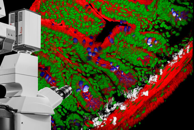
Entdecken Sie eine umfassende Sammlung wissenschaftlicher und klinischer Ressourcen, die speziell für Ärzte im Gesundheitswesen entwickelt wurden, darunter Berichte von Kollegen, klinische Fallstudien und Symposien. Speziell für Neurochirurgen, Augenärzte, plastische und rekonstruktive Chirurgen, HNO-Ärzte und Zahnärzte. Diese Sammlung präsentiert die neuesten Fortschritte in der chirurgischen Mikroskopie. Entdecken Sie, wie modernste chirurgische Technologien wie AR-Fluoreszenz, 3D-Visualisierung und intraoperative OCT-Bildgebung eine sichere Entscheidungsfindung und Präzision bei komplexen Eingriffen ermöglichen.

Fluorescence Lifetime-based Imaging Gallery
Confocal microscopy relies on the effective excitation of fluorescence probes and the efficient collection of photons emitted from the fluorescence process. One aspect of fluorescence is the emission…

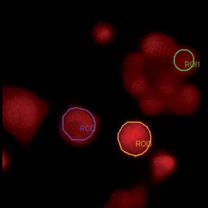
How to Quantify Changes in the Metabolic Status of Single Cells
Metabolic imaging based on fluorescence lifetime provides insights into the metabolic dynamics of cells, but its use has been limited as expertise in advanced microscopy techniques was needed.
Now,…

How FLIM Microscopy Helps to Detect Microplastic Pollution
The use of autofluorescence in biological samples is a widely used method to gain detailed knowledge about systems or organisms. This property is not only found in biological systems, but also…

Mit LIGHTNING das Maximum an Informationen aus Ihrer Probe erhalten
LIGHTNING ist ein adaptiver Prozess zur Extraktion von Bildinformationen, bei dem vollautomatisch anderweitig nicht sichtbare Strukturen und feine Details sichtbar gemacht werden. Im Gegensatz zu…

Microscopy in Virology
The coronavirus SARS-CoV-2, causing the Covid-19 disease effects our world in all aspects. Research to find immunization and treatment methods, in other words to fight this virus, gained highest…

TauSense Technology Imaging Tools
Leica Microsystems’ TauSense technology is a set of imaging modes based on fluorescence lifetime. Found at the core of the STELLARIS confocal platform, it will revolutionize your imaging experiments.…
DIVE Multiphoton Microscope Image Gallery
Today’s life science research focusses on complex biological processes, such as the causes of cancer and other human diseases. A deep look into tissues and living specimens is vital to understanding…

Step by Step Guide for FRAP Experiments
Fluorescence Recovery After Photobleaching (FRAP) has been considered the most widely applied method for observing translational diffusion processes of macromolecules. The resulting information can be…
Widefield Calcium Imaging with Calcium Indicator Fura2
In eukaryotic cells Ca2+ is one of the most widespread second messengers used in signal transduction pathways. Intracellular levels of Ca2+ are usually kept low, as Ca2+ often forms insoluble…
