
Science Lab
Science Lab
Das Wissensportal von Leica Microsystems bietet Ihnen Wissens- und Lehrmaterial zu den Themen der Mikroskopie. Die Inhalte sind so konzipiert, dass sie Einsteiger, erfahrene Praktiker und Wissenschaftler gleichermaßen bei ihrem alltäglichen Vorgehen und Experimenten unterstützen. Entdecken Sie interaktive Tutorials und Anwendungsberichte, erfahren Sie mehr über die Grundlagen der Mikroskopie und High-End-Technologien - werden Sie Teil der Science Lab Community und teilen Sie Ihr Wissen!
Loading...

RNA Quality after Different Tissue Sample Preparation
The influence of sample preparation and ultraviolet (UV) laser microdissection (UV LMD) on the quality of RNA from murine-brain tissue cryo-sections is described in this article. To obtain good…
Loading...

Digitalisierung in der neurochirurgischen Planung und bei Eingriffen
Erfahren Sie mehr über Augmented Reality, Virtual Reality und Mixed Reality in der Neurochirurgie und wie sie dazu beitragen können, Herausforderungen zu meistern.
Loading...

Augmented Reality Assisted Navigation in Neuro-Oncological Surgery
In neuro-oncological surgery, new technologies such as Augmented Reality are helping to improve surgical precision enabling a precise trajectory, conformational resection, the absence of collateral…
Loading...
Factors to Consider for a Cleanliness Analysis Solution
Choosing the right cleanliness analysis solution is important for optimal quality control. This article discusses the important factors that should be taken into account to find the solution that best…
Loading...

New Imaging Tools for Cryo-Light Microscopy
New cryo-light microscopy techniques like LIGHTNING and TauSense fluorescence lifetime-based tools reveal structures for cryo-electron microscopy.
Loading...

H&E Staining in Microscopy
If we consider the role of microscopy in pathologists’ daily routines, we often think of the diagnosis. While microscopes indeed play a crucial role at this stage of the pathology lab workflow, they…
Loading...
, the cis-golgi matrix protein GM130 (AF488, green), and the trans-golgi network membrane protein TGN46 (AF647, red).")
Golgi Organizational Changes in Response to Cell Stress
VIDEO ON DEMAND - In this episode of MicaCam, our special guest George Galea from EMBL Heidelberg will look at HeLa Kyoto cells treated with various chemotherapeutic agents to investigate their effect…
Loading...

Cleanliness Analysis for Particulate Contamination
Devices, products, and their components fabricated in many industries can be quite sensitive to contamination and, as a result, have stringent requirements for technical cleanliness. Measurement…
Loading...
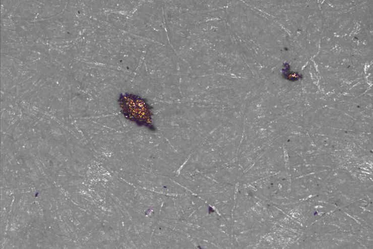
![[Translate to German:] Particles and fibers on a filter which will be counted and analyzed for cleanliness](/fileadmin/academy/2022/Efficient_particle_counting_and_analysis/Particles_and_fibers_on_a_filter.jpg "[Translate to German:] Particles and fibers on a filter which will be counted and analyzed for cleanliness")
Effiziente Partikelzählung und -analyse
Dieser Bericht befasst sich mit der Partikelzählung und -analyse unter Verwendung der optischen Mikroskopie bei der technischen Sauberkeitsanalyse von Teilen und Komponenten. Die Partikelzählung und…