
Science Lab
Science Lab
Das Wissensportal von Leica Microsystems bietet Ihnen Wissens- und Lehrmaterial zu den Themen der Mikroskopie. Die Inhalte sind so konzipiert, dass sie Einsteiger, erfahrene Praktiker und Wissenschaftler gleichermaßen bei ihrem alltäglichen Vorgehen und Experimenten unterstützen. Entdecken Sie interaktive Tutorials und Anwendungsberichte, erfahren Sie mehr über die Grundlagen der Mikroskopie und High-End-Technologien - werden Sie Teil der Science Lab Community und teilen Sie Ihr Wissen!
Loading...

How Industrial Applications Benefit from Fluorescence Microscopy
Watch this free webinar to know more about what you can do with fluorescence microscopy for industrial applications. We will cover a wide range of investigations where fluorescence contrast offers new…
Loading...
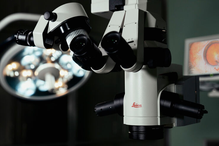
So finden Sie das richtige Mikroskop für Katarakt OPs
Vor der Auswahl eines Augenmikroskops für Kataraktoperationen und andere Eingriffe am vorderen Augenabschnitt ist es sinnvoll, sich mit der Einrichtung der Praxis oder Klinik sowie den jeweiligen…
Loading...

Intravital Microscopy of Cancer
Join our guest speaker Prof Dr Jacco van Rheenen, as he presents his work on the identity, behavior and fate of cells that drive the initiation and progression of cancer.
Loading...

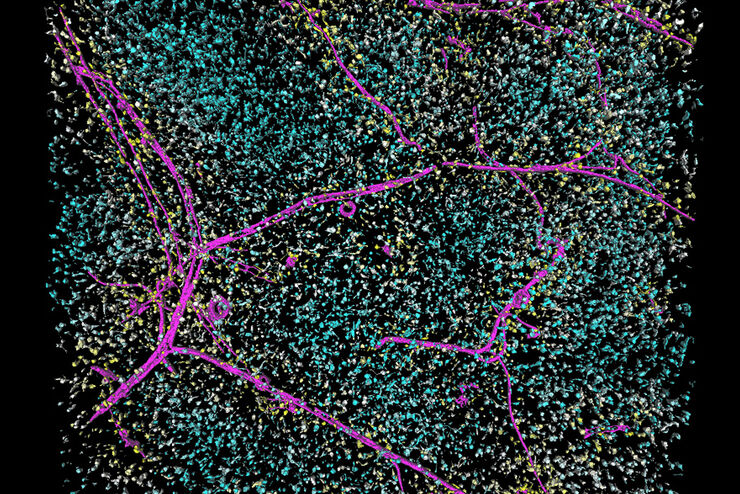
Accelerating Neuron Image Analysis with Automation
The ability to examine complex neural processes relies on the accurate reconstruction of neuronal networks at scale. Most data extraction methods in neuroscience research are time-consuming and…
Loading...
Save Time and Effort with AI-assisted Fluorescence Image Analysis
The powerful synergy of THUNDER and Aivia analyze fluorescence images with greater accuracy, even when using low light excitation.
Loading...
Tracking Single Cells Using Deep Learning
AI-based solutions continue to gain ground in the field of microscopy. From automated object classification to virtual staining, machine and deep learning technologies are powering scientific…
Loading...

Learning the Cellular Architecture from its Optical Properties
In the last 3 years, microscopists have started to use "AI based" solutions for a wide range of applications, including image acquisition optimization (smart microscopy), object classification, image…
Loading...
![[Translate to German:] Routine inspection microscope Ivesta 3](/fileadmin/_processed_/a/a/csm_Ivesta_3_integrated_monitor_users_2_fc7f6bf183.jpg "[Translate to German:] Routine inspection microscope Ivesta 3")
How to select the right solution for visual inspection
This article helps users with the decision-making process when selecting a microscope as a solution for routine visual inspection. Important factors that should be considered are described.
Loading...

How to Use a Digital Microscope to Streamline Inspection Processes
Watch this webinar for inspiration and expert advice on how to make quality control simpler, quicker, and easier. Learn how to perform comprehensive visual inspection, including comparison,…