
Science Lab
Science Lab
Das Wissensportal von Leica Microsystems bietet Ihnen Wissens- und Lehrmaterial zu den Themen der Mikroskopie. Die Inhalte sind so konzipiert, dass sie Einsteiger, erfahrene Praktiker und Wissenschaftler gleichermaßen bei ihrem alltäglichen Vorgehen und Experimenten unterstützen. Entdecken Sie interaktive Tutorials und Anwendungsberichte, erfahren Sie mehr über die Grundlagen der Mikroskopie und High-End-Technologien - werden Sie Teil der Science Lab Community und teilen Sie Ihr Wissen!
Loading...
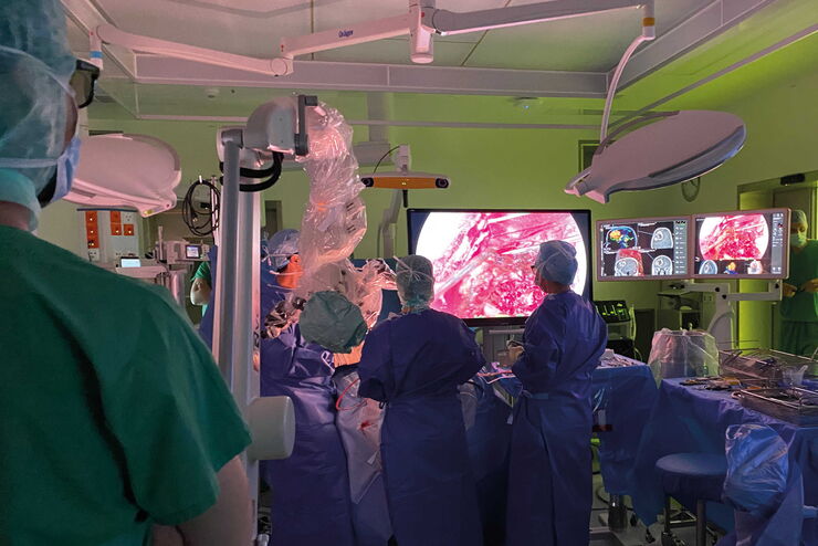
How Augmented Reality is Transforming Vascular Neurosurgery
Augmented Reality is changing surgery, with new information helping to improve the precision and safety of procedures. This is especially true in vascular neurosurgery where Augmented Reality is…
Loading...
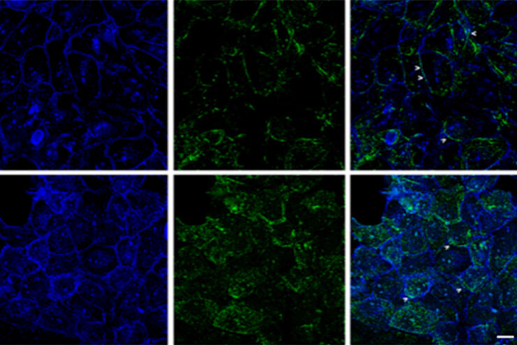
 and cytokinesis ring (Rlc1-mCherry; red).")
Studying Cell Division
Cell division is a biological process during which all cellular components must be distributed among the daughter cells. The division process requires firm coordination for success. Microscopy is…
Loading...
Improvement of Imaging Techniques to Understand Organelle Membrane Cell Dynamics
Understanding cell functions in normal and tumorous tissue is a key factor in advancing research of potential treatment strategies and understanding why some treatments might fail. Single-cell…
Loading...
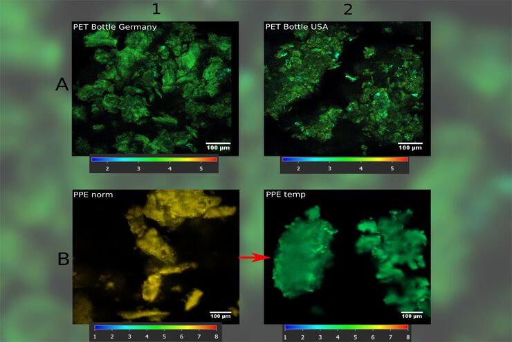
How FLIM Microscopy Helps to Detect Microplastic Pollution
The use of autofluorescence in biological samples is a widely used method to gain detailed knowledge about systems or organisms. This property is not only found in biological systems, but also…
Loading...
Oncological Reconstructive Surgery with the Leica M530 OHX Microscope
Precision is essential in oncological reconstructive surgery, in particular when it relies on free flap techniques. Microsurgical microscopes provide optimal visualization and help streamline the…
Loading...
Oncological Reconstructive Surgery: Why Use a Microscope
Recent advances in microsurgery are enhancing breast reconstruction for oncology patients, allowing both functional and aesthetic rehabilitation. More and more surgeons are adopting surgical…
Loading...
Neurosurgery with Heads-up Display
In the following video interviews Prof. Dr. Raphael Guzman, Vice Chairman of the Department of Neurosurgery at the University Hospital in Basel, Switzerland, talks about his experience in heads-up…
Loading...
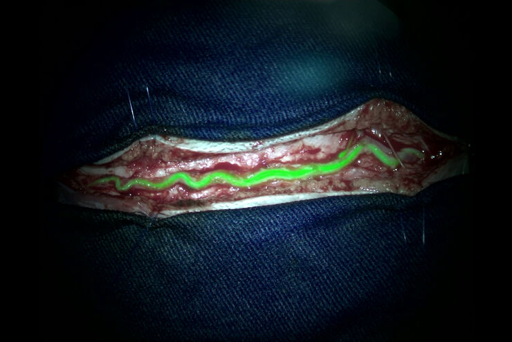
Augmented Reality (AR) Fluorescence Image Gallery
Building on a decade of leadership in fluorescence imaging technology, GLOW800 AR fluorescence is the first of many modalities based on the proprietary GLOW AR platform.
The sophisticated imaging…
Loading...
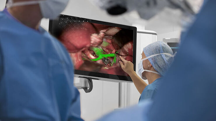
Clinical Uses in Cerebrovascular and Skull Base Neurosurgery
In this webinar Dr. Bendok and Dr. Morcos explain how Augmented Reality and Fluorescence can enhance visualization and support surgical decision making. They present first-hand experience of the GLOW…