

Science Lab
Science Lab
Bienvenue sur le portail de connaissances de Leica Microsystems. Vous y trouverez des recherches scientifiques et du matériel didactique sur le thème de la microscopie. Le portail aide les débutants, les praticiens expérimentés et les scientifiques dans leur travail quotidien et leurs expériences. Explorez les didacticiels interactifs et les notes d'application, découvrez les bases de la microscopie ainsi que les technologies de pointe. Faites partie de la communauté Science Lab et partagez votre expertise.
Filter articles
Tags
Type de publication
Produits
Loading...
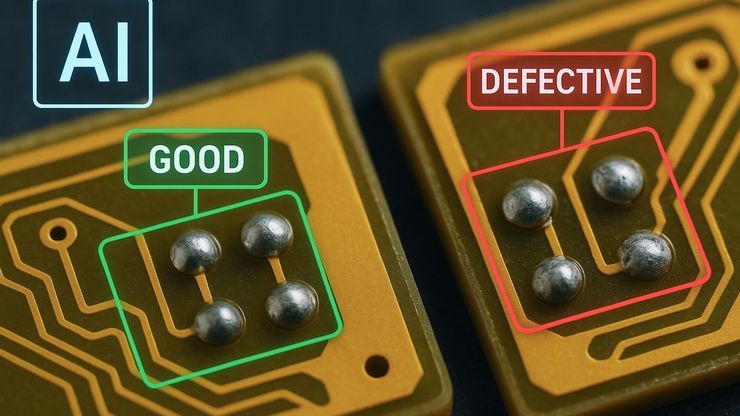
AI Image Analysis for Reliable Defect Detection
AI-based visual inspection with microscopy helps make quality control in industrial environments efficient by reducing manual inspection effort concerning defect detection, classification, and…
Loading...

An Overview of Artificial Intelligence (AI) and Machine Learning
AI is rapidly changing industrial inspection, quality control, and manufacturing workflows. But not all AI technologies work the same way. From machine learning and deep learning to the newer concept…
Loading...

Why the Future of Microscopic Inspection Is AI-Assisted, Not Fully Automated
The next wave of QC productivity gains will not come from building more automated inspection lines. Instead, it will come from augmenting millions of inspection decisions that are made manually every…
Loading...

How to Select the Right Measurement Microscope
With a measurement microscope, users can measure the size and dimensions of sample features in both 2D and 3D, something crucial for inspection, QC, failure analysis, and R&D. However, choosing the…
Loading...
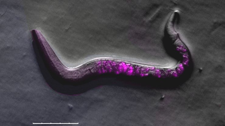
A Guide to C. elegans Research – Working with Nematodes
Efficient microscopy techniques for C. elegans research are outlined in this guide. As a widely used model organism with about 70% gene homology to humans, the nematode Caenorhabditis elegans (also…
Loading...
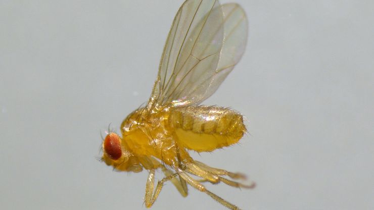
A Guide to Using Microscopy for Drosophila (Fruit Fly) Research
The fruit fly, typically Drosophila melanogaster, has been used as a model organism for over a century. One reason is that many disease-related genes are shared between Drosophila and humans. It is…
Loading...
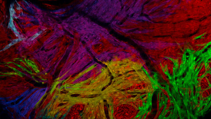
A Guide to Zebrafish Research
To obtain optimal results while doing zebrafish research, especially during screening, sorting, handling, and imaging, seeing the fine details and structures is important. They help researchers make…
Loading...
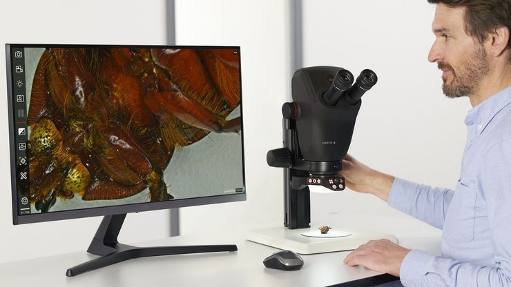
Microscopes de dissection
Si vous devez procéder à des dissections, il peut vous arriver de rester penché sur les oculaires du microscope pendant des heures. Leica Microsystems vous propose de faire votre choix parmi un large…
Loading...
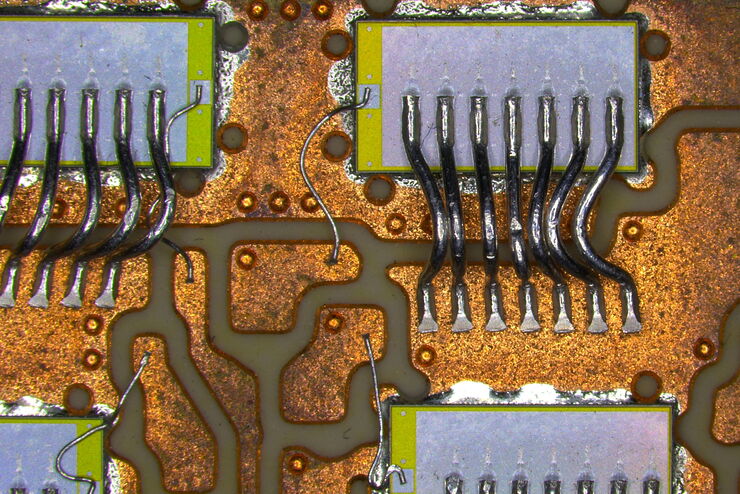
Top Challenges for Visual Inspection
This article discusses the challenges encountered when performing visual inspection and rework using a microscope. Using the right type of microscope and optical setup is paramount in order to…


