
Science Lab
Science Lab
Bienvenue sur le portail de connaissances de Leica Microsystems. Vous y trouverez des recherches scientifiques et du matériel didactique sur le thème de la microscopie. Le portail aide les débutants, les praticiens expérimentés et les scientifiques dans leur travail quotidien et leurs expériences. Explorez les didacticiels interactifs et les notes d'application, découvrez les bases de la microscopie ainsi que les technologies de pointe. Faites partie de la communauté Science Lab et partagez votre expertise.
Filter articles
Tags
Type de publication
Produits
Loading...

Computational Clearing - Enhance 3D Specimen Imaging
This webinar is designed to clarify crucial specifications that contribute to THUNDER Imagers' transformative visualization of 3D samples and improvements within a researcher's imaging-related…
Loading...

Overcoming Complexities in Microdentistry
Dr. Salam Abu Arqub, from the Smile Engineer Dental Center in Amman, Jordan, has been using Leica dental microscopes for three years for all procedures performed at the clinic. He shared his…
Loading...

THUNDER Imagers: High Performance, Versatility and Ease-of-Use for your Everyday Imaging Workflows
This webinar will showcase the versatility and performance of THUNDER Imagers in many different life science applications: from counting nuclei in retina sections and RNA molecules in cancer tissue…
Loading...
Digital Classroom Options
As teachers, you know your big challenge is to catch and keep the students’ attention and the best chance for this is by making the environment interactive. In the case of the Microscopy Classroom, we…
Loading...
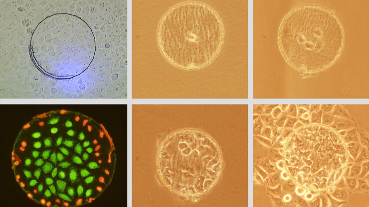
Live Cell Isolation by Laser Microdissection
Laser microdissection is a tool for the isolation of homogenous cell populations from their native niches in tissues to downstream molecular assays. Beside its routine use for fixed tissue sections,…
Loading...
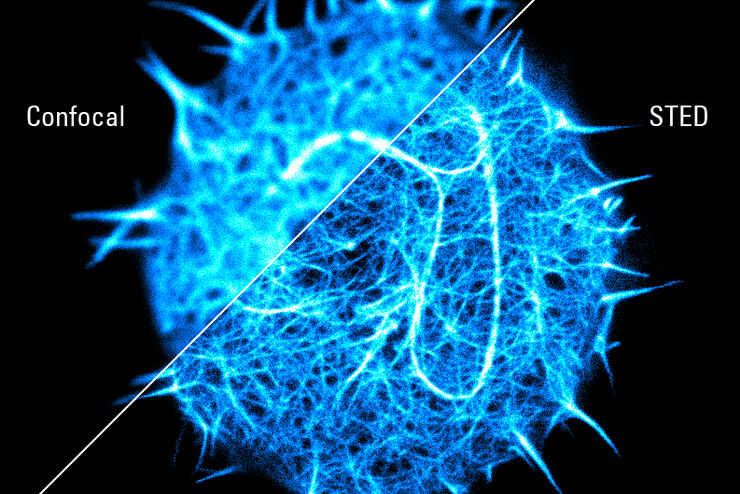
Super-resolved STED spectroscopy
Molecular interactions are key in cellular signalling. They are often ruled or rendered by the mobility of the involved molecules.
Loading...
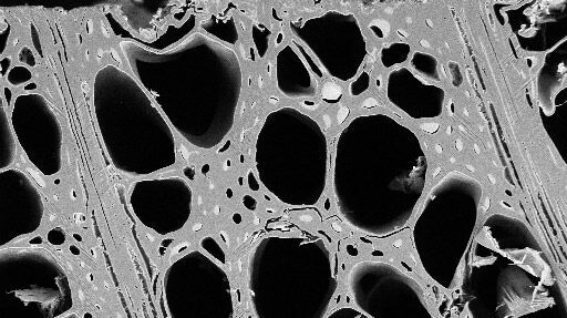
Practical Applications of Broad Ion Beam Milling
Mechanical polishing can be time consuming and frustrating. It can also introduce unwanted artifacts when preparing cross-sectioned samples for electron backscatter diffraction (EBSD) in the scanning…
Loading...

Digital Microscopy in Earth Science
Classical polarized light (compound) microscopes can only be used for prepared samples, because the working distance they offer is insufficient for whole samples. This means that thicker and bigger…
