
Industrial Microscopy
Industrial Microscopy
Dive deep into detailed articles and webinars focusing on efficient inspection, optimized workflows, and ergonomic comfort in industrial and pathological contexts. Topics covered include quality control, materials analysis, microscopy in pathology, among many others. This is the place where you get valuable insights into using cutting-edge technologies for improving precision and efficiency in manufacturing processes as well as accurate pathological diagnosis and research.

Microscopy in Virology
The coronavirus SARS-CoV-2, causing the Covid-19 disease effects our world in all aspects. Research to find immunization and treatment methods, in other words to fight this virus, gained highest…

Top Issues Related to Standards for Rating Non-Metallic Inclusions in Steel
Supplying components and products made of steel to users worldwide can require that a single batch be compliant with multiple steel quality standards. This user demand creates significant challenges…

Analyzing Non-metallic Inclusions in Steel
Oftentimes we find ourselves caught up in tedious analyses by reticle and comparison chart, time-consuming double-evaluation according to several standards or subjective inspection results with a bias…

Rate the Quality of Your Steel: Free Webinar and Report
This webinar and report describe optimal microscopy solutions for rating steel quality in terms of non-metallic inclusions and reviews the various international and regional standards concerning…
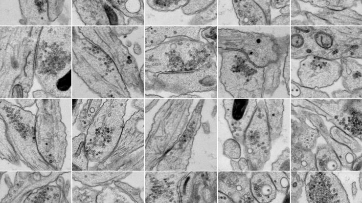
Bridging Structure and Dynamics at the Nanoscale through Optogenetics and Electrical Stimulation
Nanoscale ultrastructural information is typically obtained by means of static imaging of a fixed and processed specimen. However, this is only a snapshot of one moment within a dynamic system in…

High Resolution Array Tomography with Automated Serial Sectioning
The optimization of high resolution, 3-dimensional (3D), sub-cellular structure analysis with array tomography using an automated serial sectioning solution, achieving a high section density on the…

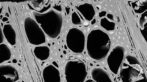
Macroscale to Nanoscale Pore Analysis of Shale and Carbonate Rocks
Physical porosity in rocks, like shale and carbonate, has a large effect on the their storage capacity. The pore geometries also affect their permeability. Imaging the visible pore space provides…
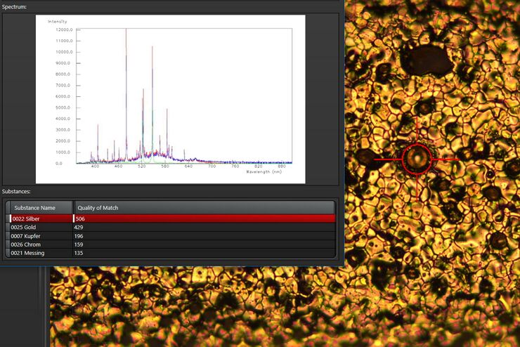
See the Structure with Microscopy - Know the Composition with Laser Spectroscopy
The advantages of a 2-in-1 materials analysis solution combining optical microscopy and laser induced breakdown spectroscopy (LIBS) for simultaneous visual and chemical inspection are described in…
Practical Applications of Broad Ion Beam Milling
Mechanical polishing can be time consuming and frustrating. It can also introduce unwanted artifacts when preparing cross-sectioned samples for electron backscatter diffraction (EBSD) in the scanning…
