
Industrial Microscopy
Industrial Microscopy
Dive deep into detailed articles and webinars focusing on efficient inspection, optimized workflows, and ergonomic comfort in industrial and pathological contexts. Topics covered include quality control, materials analysis, microscopy in pathology, among many others. This is the place where you get valuable insights into using cutting-edge technologies for improving precision and efficiency in manufacturing processes as well as accurate pathological diagnosis and research.

Immersion Freezing for Cryo-Transmission Electron Microscopy: Applications
A well established usage case for cryo-TEM is three-dimensional reconstruction of isolated macromolecules, virus particles, or filaments. These approaches are based on averaging of repetitive…
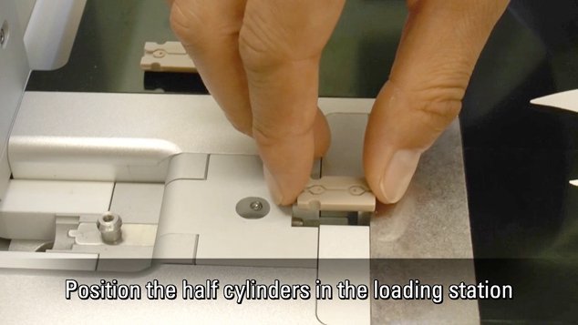
Video Tutorials: Filling and Assembling of Different Carriers for High-Pressure Freezing
High pressure freezing (HPF) is a cryo-fixation method primarily for biological samples, but also for a variety of non-biological materials. It is a technique that yields optimal preservation in many…
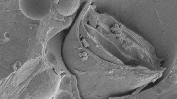
Brief Introduction to Freeze Fracture and Etching
Freeze fracture describes the technique of breaking a frozen specimen to reveal internal structures. Freeze etching is the sublimation of surface ice under vacuum to reveal details of the fractured…
Brief Introduction to Specimen Trimming
Before ultrathin sectioning a sample with an ultramicrotome it has to be pre-prepared. For this pre-preparation, special attention must be paid to the sample size (size of the section), location of…
Glycerol Spraying/Platinum Low Angle Rotary Shadowing of DNA with the Leica EM ACE600 e-beam
Glycerol spraying/low angle rotary shadowing (Aebi and Baschong, 2006) is a preparation technique used in biology to visualize structures yielding insufficent contrast with other techniques, due to…

Brief Introduction to Freeze Substitution
Freeze-substitution is a process of dehydration, performed at temperatures low enough to avoid the formation of ice crystals and to circumvent the damaging effects observed after ambient-temperature…
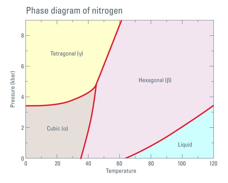
Thermodynamic Considerations Regarding the LN2 in a High Pressure Freezer
Employing liquid nitrogen (LN2) as a coolant in the complex process of high pressure freezing raises certain considerations regarding phase transition not only of the liquid sample to be frozen but…

Brief Introduction to Contrasting for EM Sample Preparation
Since the contrast in the electron microscope depends primarily on the differences in the electron density of the organic molecules in the cell, the efficiency of a stain is determined by the atomic…

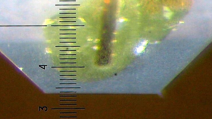
Brief Introduction to Glass Knifemaking for Electron and Light Microscope Applications
Glass knives are used in an ultramicrotome to cut ultrathin slices of samples for electron and light microscope applications. For resin and for cryo sections (Tokuyasu samples) the knife edge must be…
