
Science Lab
Science Lab
Das Wissensportal von Leica Microsystems bietet Ihnen Wissens- und Lehrmaterial zu den Themen der Mikroskopie. Die Inhalte sind so konzipiert, dass sie Einsteiger, erfahrene Praktiker und Wissenschaftler gleichermaßen bei ihrem alltäglichen Vorgehen und Experimenten unterstützen. Entdecken Sie interaktive Tutorials und Anwendungsberichte, erfahren Sie mehr über die Grundlagen der Mikroskopie und High-End-Technologien - werden Sie Teil der Science Lab Community und teilen Sie Ihr Wissen!
Loading...

Studying Ocular Birth Defects
This article discusses how lens formation and ocular birth defects can be studied with sharp widefield microscopy images which are acquired rapidly. The mouse ocular lens is used as a model to study…
Loading...

Studying Wound Healing of Smooth Muscle Cells
This article discusses how wound healing of cultured smooth muscle cells (SMCs) in multiwell plates can be reliably studied over time with less effort using a specially configured Leica inverted…
Loading...
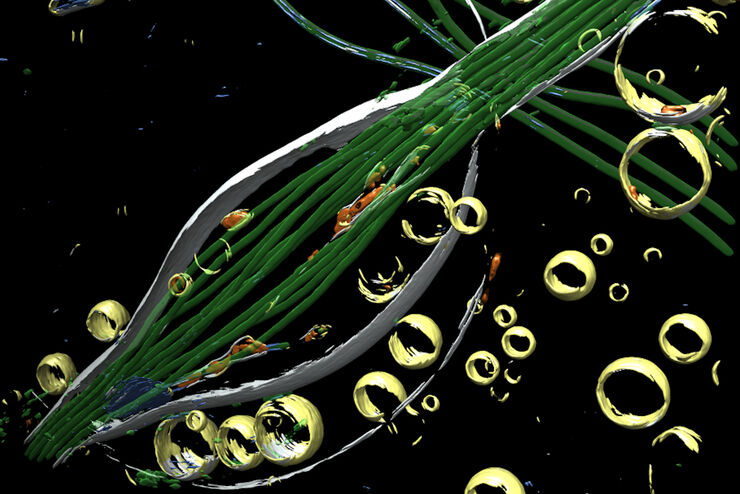
 and mito OM (red) in a live U2OS cell")
Multicolor 4D Super Resolution Light Sheet Microscopy
The AI Microscopy Symposium offers a unique forum for discussing the latest AI-based technologies and tools in the field of microscopy and biomedical imaging. In this scientific presentation, Yuxuan…
Loading...
 and THUNDER (right) image of Ewing Sarcoma cells (SK-ES-1)")
Visualizing the Mitotic Spindle in Cancer Cells
This article demonstrates how this research is aided by visualizing more details of mitotic spindles in Ewing Sarcoma cells using the THUNDER Imager Tissue and Large Volume Computational Clearing…
Loading...

How to Keep Your Samples Under Physiological Conditions
The Coral Life workflow combines dynamic data with the best possible sample fixation by high pressure freezing. However, good sample preservation won’t help if your cells are stressed by temperature…
Loading...
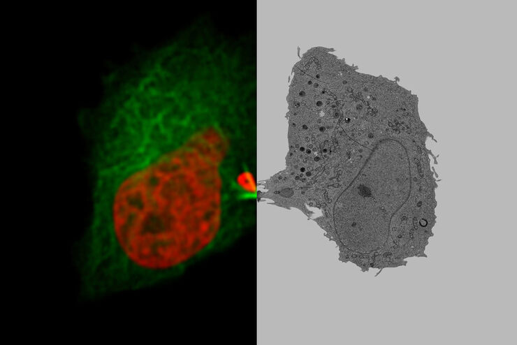
Putting Dynamic Live Cell Data into the Ultrastructural Context
With workflow Coral Life, searching for a needle in the haystack is a thing of the past. Take advantage of correlative light and electron microscopy to identify directly the right cell at the right…
Loading...
Advancing Cell Biology with Cryo-Correlative Microscopy
Correlative light and electron microscopy (CLEM) advances biological discoveries by merging different microscopes and imaging modalities to study systems in 4D. Combining fluorescence microscopy with…
Loading...

Development of Fluorescence Lifetime Imaging Microscopy (FLIM) and its Relevance for Functional Imaging
Prof. Ammasi Periasamy, Director, Keck Center for Cellular Imaging, University of Virginia, was interviewed by Dr. Giulia Ossato, Product Manager functional imaging, during Leica Microsystems Meets…
Loading...
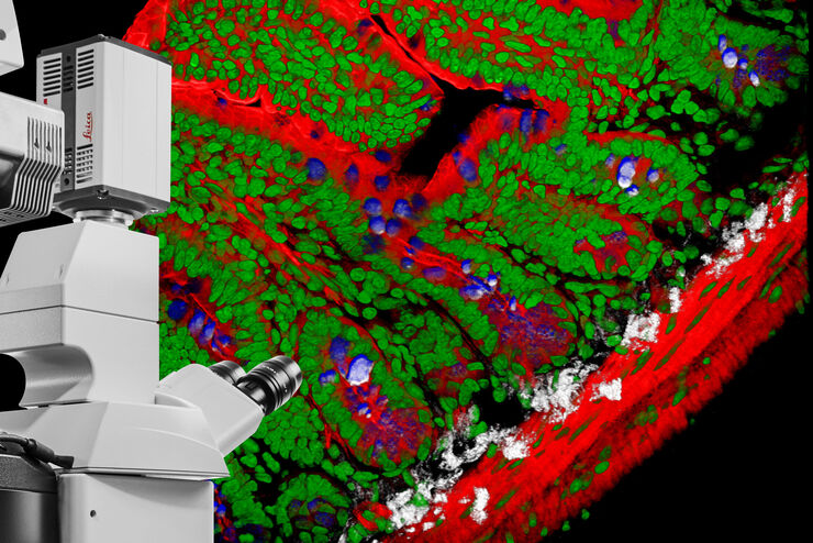
DIVE Multiphoton Microscope Image Gallery
Today’s life science research focusses on complex biological processes, such as the causes of cancer and other human diseases. A deep look into tissues and living specimens is vital to understanding…