
Science Lab
Science Lab
Das Wissensportal von Leica Microsystems bietet Ihnen Wissens- und Lehrmaterial zu den Themen der Mikroskopie. Die Inhalte sind so konzipiert, dass sie Einsteiger, erfahrene Praktiker und Wissenschaftler gleichermaßen bei ihrem alltäglichen Vorgehen und Experimenten unterstützen. Entdecken Sie interaktive Tutorials und Anwendungsberichte, erfahren Sie mehr über die Grundlagen der Mikroskopie und High-End-Technologien - werden Sie Teil der Science Lab Community und teilen Sie Ihr Wissen!
Loading...
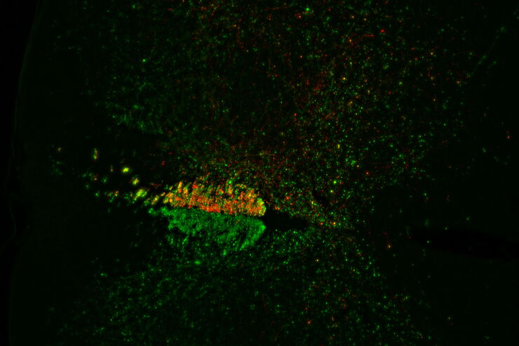
Evaluating Axon Regeneration After Brain or Spine Trauma of Mice
Damaged nerve regeneration was investigated using mouse spinal cord sections treated with compounds that counter axon growth inhibitor (AGI) proteins. The sections were screened to find active and…
Loading...
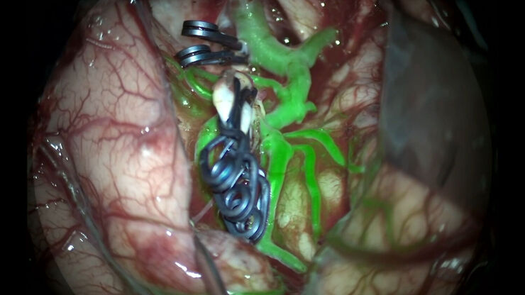
GLOW800 Augmented Reality Fluorescence in Aneurysm Treatment
This case study from Prof. Dr. Feres Chaddad talks about the treatment of unruptured MCA (middle cerebral artery) and PCOM (posterior communicating artery) aneurysms with microsurgical clipping. It…
Loading...
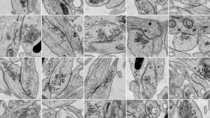
Bridging Structure and Dynamics at the Nanoscale through Optogenetics and Electrical Stimulation
Nanoscale ultrastructural information is typically obtained by means of static imaging of a fixed and processed specimen. However, this is only a snapshot of one moment within a dynamic system in…
Loading...

Zebrafish Brain - Whole Organ Imaging at High Resolution
Structural information is key when one seeks to understand complex biological systems, and one of the most complex biological structures is the vertebrate central nervous system. To image a complete…
Loading...
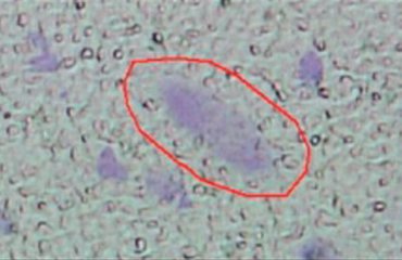
Improving RNA Analysis with Laser Microdissection
Parkinson’s disease is a progressive neurodegenerative disorder connected with cell death of dopamine-releasing neurons in the brain. Differences in gene expression between individual…
Loading...
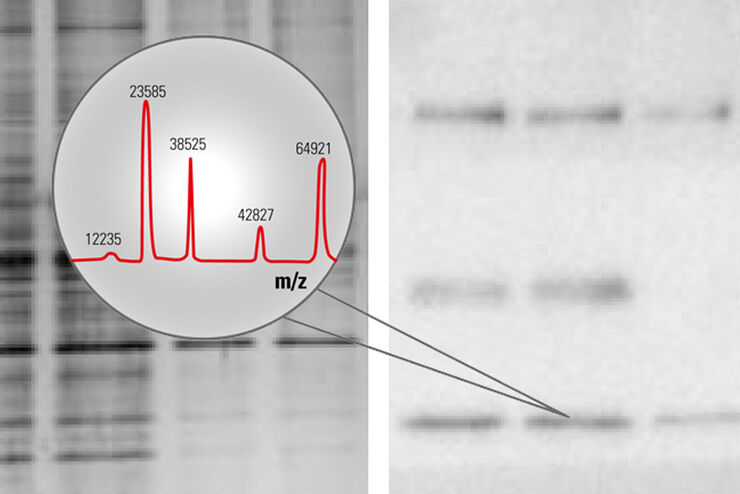
How to improve your Alzheimer Protein Analysis with Laser Microdissection
Brain Research: Collect pure starting material for proteomics - Improve your workflow with Laser Microdissection - Many brain diseases result from protein malfunction, misfolding and agglutination.…
Loading...

Studying Caenorhabditis elegans (C. elegans)
Find out how you can image and study C. elegans roundworm model organisms efficiently with a microscope for developmental biology applications from this article.
Loading...
The Morbus Parkinson Puzzle
A characteristic sign of M. Parkinson is the deterioration of dopaminergic neurons in the mid-brain, specifically in the substantia nigra (SN, black substance). Different causes and forms of this…
Loading...

Ratiometric Imaging
Many fundamental functions of a cell strongly depend on delicate, but nevertheless dynamic balances of ions (e.g. calcium, magnesium), voltage potentials and pH between the cell’s cytosol and the…