
Science Lab
Science Lab
Das Wissensportal von Leica Microsystems bietet Ihnen Wissens- und Lehrmaterial zu den Themen der Mikroskopie. Die Inhalte sind so konzipiert, dass sie Einsteiger, erfahrene Praktiker und Wissenschaftler gleichermaßen bei ihrem alltäglichen Vorgehen und Experimenten unterstützen. Entdecken Sie interaktive Tutorials und Anwendungsberichte, erfahren Sie mehr über die Grundlagen der Mikroskopie und High-End-Technologien - werden Sie Teil der Science Lab Community und teilen Sie Ihr Wissen!
Loading...
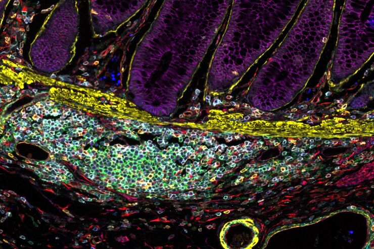
Designing your Research Study with Multiplexed IF Imaging
Multiplexed tissue analysis is a powerful technique that allows comparisons of cell-type locations and cell-type interactions within a single fixed tissue sample. It is common for researchers to ask…
Loading...
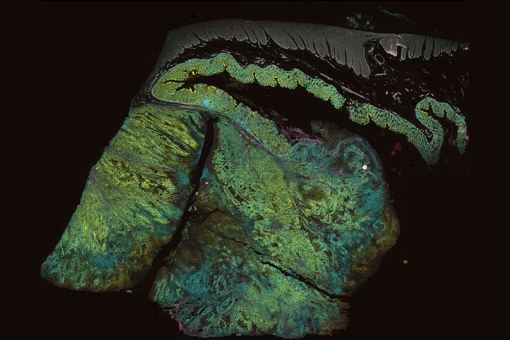
Be Confident in your Results with Cell DIVE Validated Antibodies
The Cell DIVE System includes a carefully curated list of hundreds of commercially available antibodies validated to offer optimal specificity and sensitivity in multiplexed imaging. That validation…
Loading...
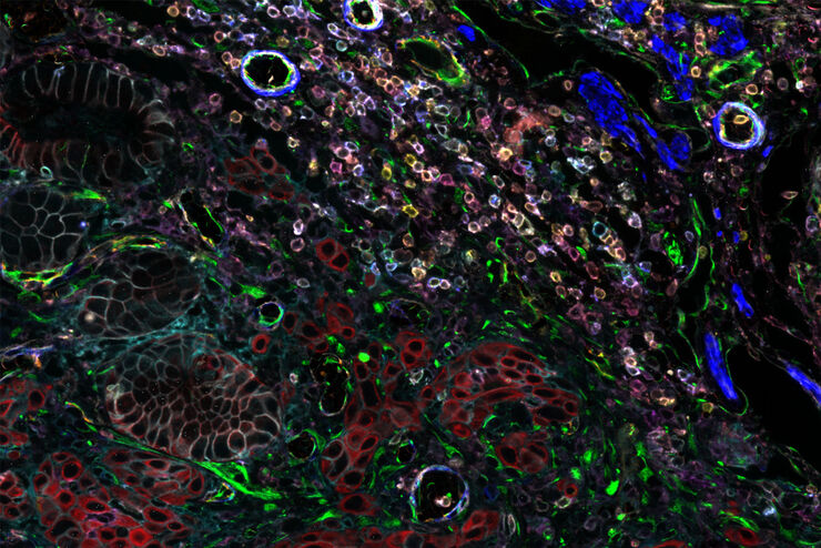
Overcoming Multiplexed Imaging Roadblocks
Whether a single slide or a batch of slides processed on multiple Cell DIVE imagers, every step carried out by the Cell DIVE Acquisition Software eliminates distortions and artifacts and seamlessly…
Loading...
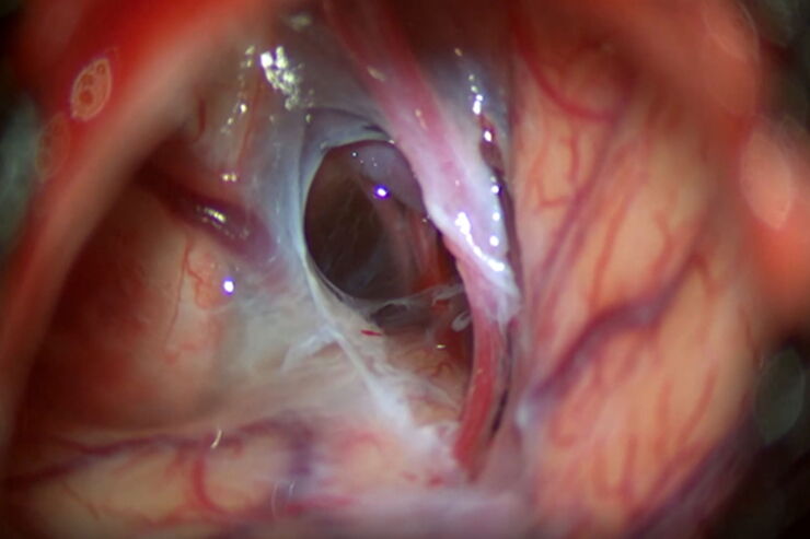
Optimal Visualization in Brain Surgery
This case study “Treatment of the Galassi type III arachnoid cyst with the M530 OHX surgical microscope from Leica Microsystems” documents the procedure step by step and shows the visualization…
Loading...
 and astrocytes (green) in a cortical spheroid derived from human induced pluripotent stem cells.")
Neuroscience Images
Neuroscience commonly uses microscopy to study the nervous system’s function and understand neurodegenerative diseases.
Loading...
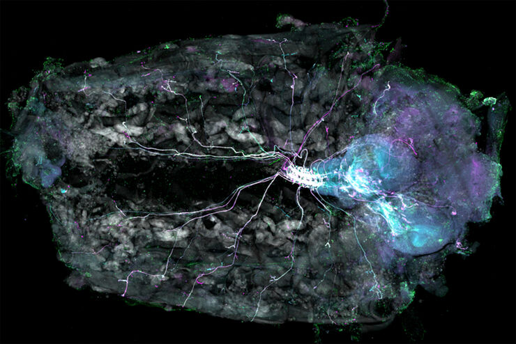
Understanding Motor Sequence Generation Across Spatiotemporal Scales
We have developed a microscopy-based pipeline to characterize a developmentally critical behavior at the pupal stage of development, called the ecdysis sequence. We study brain-wide neuronal activity…
Loading...
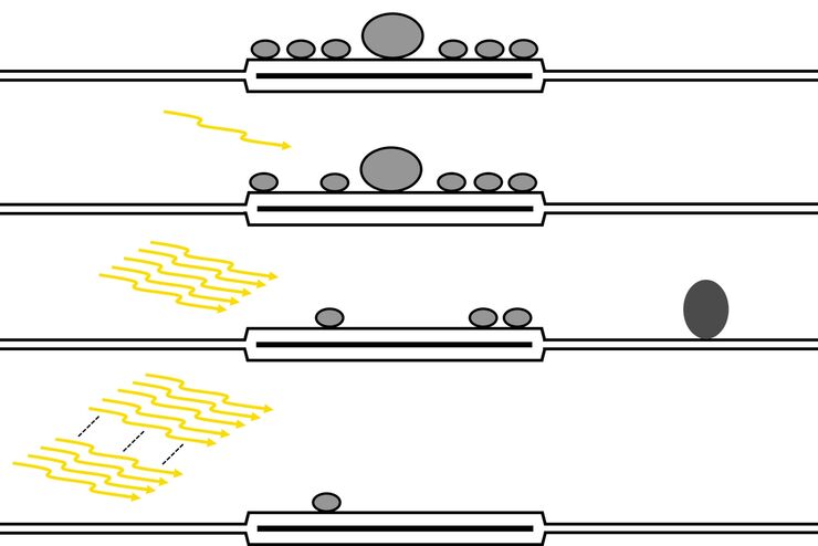
Investigating Synapses in Brain Slices with Enhanced Functional Electron Microscopy
A fundamental question of neuroscience is: what is the relationship between structural and functional properties of synapses? Over the last few decades, electrophysiology has shed light on synaptic…
Loading...
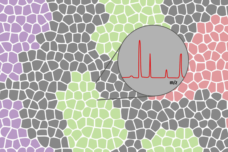
How to improve your Biomarker Discovery Workflow with Laser Microdissection
Biomarkers can be used as indicators of certain diseases, such as cancer. The tumor microenvironment moved into the spotlight in this concern. It is in close interaction with the tumor itself.…
Loading...
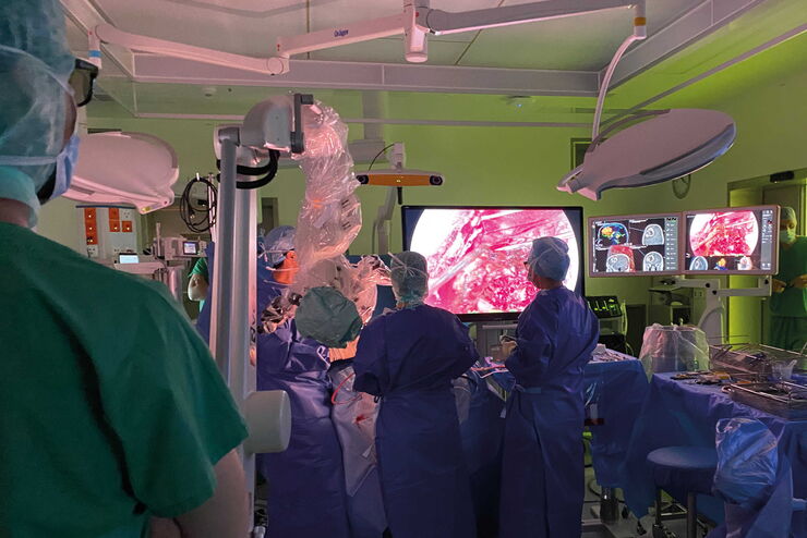
Neurosurgery with Heads-up Display
In the following video interviews Prof. Dr. Raphael Guzman, Vice Chairman of the Department of Neurosurgery at the University Hospital in Basel, Switzerland, talks about his experience in heads-up…