
Science Lab
Science Lab
Bienvenido al portal de conocimiento de Leica Microsystems. Aquí encontrará investigación científica y material didáctico sobre el tema de la microscopía. El portal ayuda a principiantes, profesionales experimentados y científicos por igual en su trabajo diario y en sus experimentos. Explore tutoriales interactivos y notas de aplicación, descubra los fundamentos de la microscopía, así como las tecnologías de gama alta. Forme parte de la comunidad Science Lab y comparta sus conocimientos.
Filter articles
Tags
Story Type
Products
Loading...

Understanding Motor Sequence Generation Across Spatiotemporal Scales
We have developed a microscopy-based pipeline to characterize a developmentally critical behavior at the pupal stage of development, called the ecdysis sequence. We study brain-wide neuronal activity…
Loading...

Benefits of TauContrast to Image Complex Samples
In this interview, Dr. Timo Zimmermann talks about his experience with the application of TauSense tools and their potential for the investigation of demanding samples such as thick samples or…
Loading...
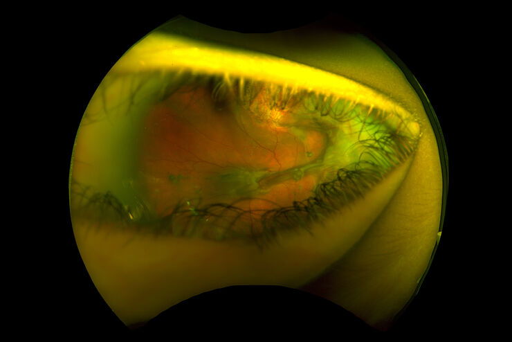
Intraoperative OCT-Assisted Surgical Management of Proliferative Vitreoretinopathy
Proliferative vitreoretinopathy (PVR) is a plague to patients and their surgeons after recent rhegmatogenous retinal detachment (RD). Despite excellent initial surgical outcomes, it is the most common…
Loading...

Fast, High-quality Vitrification with the EM ICE High Pressure Freezer
The EM ICE High Pressure Freezer was developed with a unique freezing principle and uses only a single pressurization and cooling liquid: liquified nitrogen (LN2). This design enables three major…
Loading...

Targeting Active Recycling Nuclear Pore Complexes using Cryo Confocal Microscopy
In this article, how cryo light microscopy and, in particular cryo confocal microscopy, is used to improve the reliability of cryo EM workflows is described. The quality of the EM grids and samples is…
Loading...
How Augmented Reality is Transforming Vascular Neurosurgery
Augmented Reality is changing surgery, with new information helping to improve the precision and safety of procedures. This is especially true in vascular neurosurgery where Augmented Reality is…
Loading...
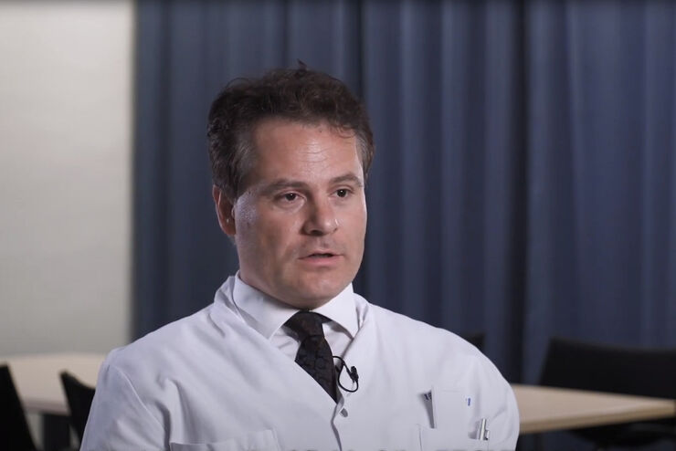
Overcoming Ophthalmologic Surgery Challenges
Ophthalmology surgical procedures involving both the anterior and posterior segment can be particularly challenging. Good visualization is required to operate with precision and confidence.
Prof.…
Loading...

Moving to Routine Use of Intraoperative OCT in Eye Surgery
Dr Barbara Parolini is one of the pioneers in the use of intraoperative OCT in eye surgery. Since 2016, she has gained a lot of clinical experience with different intraoperative OCT systems. In this…
Loading...

Investigating Synapses in Brain Slices with Enhanced Functional Electron Microscopy
A fundamental question of neuroscience is: what is the relationship between structural and functional properties of synapses? Over the last few decades, electrophysiology has shed light on synaptic…
