
Science Lab
Science Lab
Bienvenue sur le portail de connaissances de Leica Microsystems. Vous y trouverez des recherches scientifiques et du matériel didactique sur le thème de la microscopie. Le portail aide les débutants, les praticiens expérimentés et les scientifiques dans leur travail quotidien et leurs expériences. Explorez les didacticiels interactifs et les notes d'application, découvrez les bases de la microscopie ainsi que les technologies de pointe. Faites partie de la communauté Science Lab et partagez votre expertise.
Filter articles
Tags
Type de publication
Produits
Loading...

Towards Advanced Use of Intraoperative OCT in Cataract Surgery
In this White Paper, Dr. Rachid Tahiri shares his personal experience with the Leica EnFocus intraoperative OCT, the valuable features supporting smooth surgery and how it allows him to minimize…
Loading...

How Clinical and Surgical Microscopes Support Otolaryngology
The number of ENT surgery procedures is increasing year after year. It is estimated that there will be over 21 million ENT procedures performed annually worldwide by 2022.
Dr. Duane Mol is the…
Loading...
 which is published by the US FDA (Food & Drug Administration).")
Introduction to 21 CFR Part 11 and Related Regulations
This article provides an overview of regulations and guidelines for electronic records (data entry, storage, signatures, and approvals) used in the USA (21 CFR Part 11), EU (GMP Annex 11), and China…
Loading...

Physiology Image Gallery
Physiology is about the processes and functions within a living organism. Research in physiology focuses on the activities and functions of an organism’s organs, tissues, or cells, including the…
Loading...

Live Cell Imaging Gallery
Live cell microscopy techniques are fundamental to get a better understanding of cellular and molecular function. Today, widefield microscopy is the most common technique used to visualize cell…
Loading...

Tissue Image Gallery
Visual analysis of animal and human tissues is critical to understand complex diseases such as cancer or neurodegeneration. From basic immunohistochemistry to intravital imaging, confocal microscopy…
Loading...
 and astrocytes (green) in a cortical spheroid derived from human induced pluripotent stem cells.")
Neuroscience Images
Neuroscience commonly uses microscopy to study the nervous system’s function and understand neurodegenerative diseases.
Loading...

Multicolor Image Gallery
Fluorescence multicolor microscopy, which is one aspect of multiplex imaging, allows for the observation and analysis of multiple elements within the same sample – each tagged with a different…
Loading...
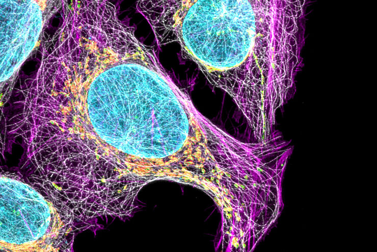
Cell Biology Image Gallery
Cell biology studies the structure, function and behavior of cells, including cell metabolism, cell cycle, and cell signaling. Fluorescence microscopes are an integral part of a cell biologist…
