Leica Microsystems

Leica Microsystems

Leica Microsystems is a world leader in microscopes and scientific instruments. Founded as a family business in the nineteenth century, the company’s history was marked by unparalleled innovation on its way to becoming a global enterprise.

Its historically close cooperation with the scientific community is the key to Leica Microsystems’ tradition of innovation, which draws on users’ ideas and creates solutions tailored to their requirements. At the global level, Leica Microsystems is organized in three divisions, all of which are among the leaders in their respective fields: Life Science, Industry and Medical.

The company is represented in over 100 countries with 6 manufacturing facilities in 5 countries, sales and service organizations in 20 countries, and an international network of dealers. The company is headquartered in Wetzlar, Germany.

http://www.leica-microsystems.com/

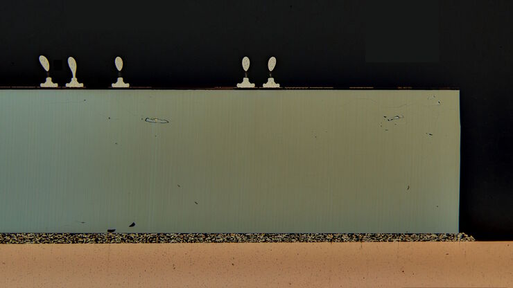
Image of an integrated-circuit (IC) chip cross section acquired at higher magnification showing a region of interest.

Structural and Chemical Analysis of IC-Chip Cross Sections

This article shows how electronic IC-chip cross sections can be efficiently and reliably prepared and then analyzed, both visually and chemically at the microscale, with the EM TXP and DM6 M LIBS
EBSD grain size distribution of the cross section of a gold wire within a silicon matrix from inside a CPU (central processing unit of a computer). The grains are highlighted with arbitrary colors.

High-Quality EBSD Sample Preparation

This article describes a method for EBSD sample preparation of challenging materials. The high-quality samples required for electron backscatter diffraction are prepared with broad ion-beam milling.
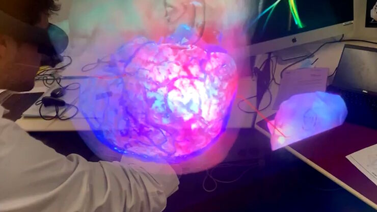
Augmented Reality fluorescence supports each step of neurovascular surgery procedures. Image courtesy of Dr. Christof Renner.

Use of AR Fluorescence in Neurovascular Surgery

Learn about the use of GLOW800 Augmented Reality in neurovascular surgery through clinical cases and videos, including aneurysm and tumor resection cases.
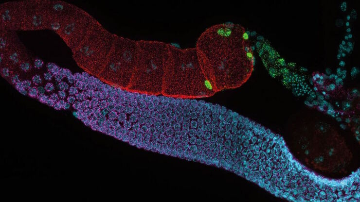
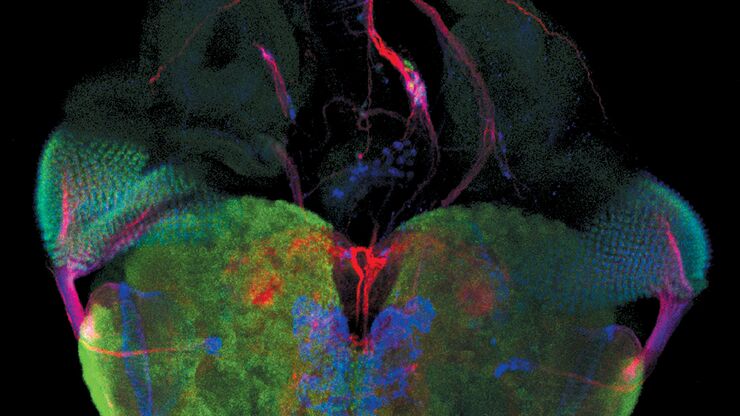
C. elegans adult hermaphrodite gonades acquired using THUNDER Imager. Staining: blue - DAPI (nucleus), green - SP56 (sperm), red - RME-2 (oocyte), magenta - PGL-1 (RNA + protein granules). Image courtesy of Prof. Dr. Christian Eckmann, Martin Luther University, Halle, Germany.
Life Science Research: Which Microscope Camera is Right for You?

Deciding which microscope camera best fits your experimental needs can be daunting. This guide presents the key factors to consider when selecting the right camera for your life science research.
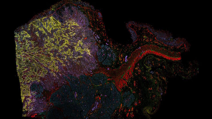
Multi-tissue array with 4 markers shown including DAPI, NaKATPase, PanCk, and Vimentin.

Spatial Biology: Learning the Landscape

Spatial Biology: Understanding the organization and interaction of molecules, cells, and tissues in their native spatial context
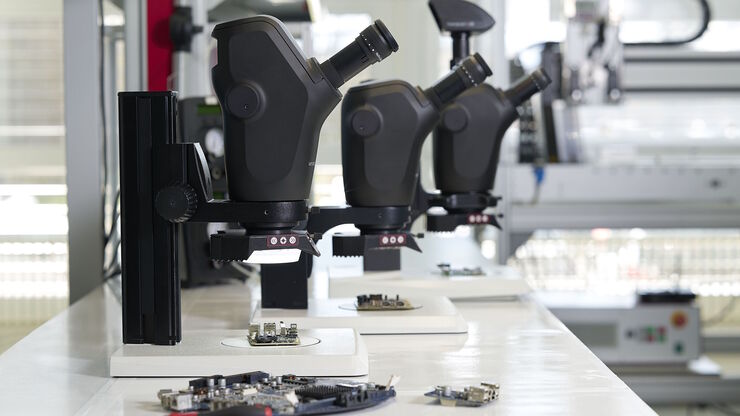
Stereo microscopes are often considered the workhorses of laboratories and production sites.

Key Factors to Consider When Selecting a Stereo Microscope

This article explains key factors that help users determine which stereo microscope solution can best meet their needs, depending on the application.
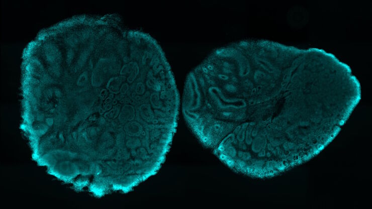
Brain organoid section (DAPI) acquired using THUNDER Imager Live Cell. Image courtesy of Janina Kaspar and Irene Santisteban, Schäfer Lab, TUM.

Imaging Organoid Models to Investigate Brain Health

Imaging human brain organoid models to study the phenotypes of specialized brain cells called microglia, and the potential applications of these organoid models in health and disease.

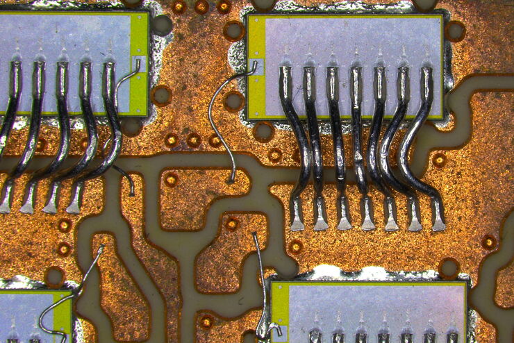
Rapid and Reliable Examination of PCBs & PCBAs with Digital Microscopy

Digital microscopes provide users with a convenient and rapid way to acquire high-quality, reliable image data and make quick inspection and analysis of printed circuit boards (PCBs) and assemblies…
In vivo imaging of a mouse pial and cortical vasculature through a glass window (ROSAmT/mG::Pdgfb-CreERT2 mouse meningeal and cortical visualization following tamoxifen induction and craniotomy). Courtesy: Thomas Mathivet, PhD

Windows on Neurovascular Pathologies

Discover how innate immunity can sustain deleterious effects following neurovascular pathologies and the technological developments enabling longitudinal studies into these events.
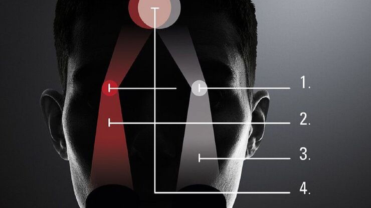
The principle of the FusionOptics technology:  Of the two separate beam paths (1), one provides depth of field (2) and the other high resolution (3). In the brain, the two images of the sample are merged into a single, optimal 3D image (4).

What is the FusionOptics Technology?

Leica stereo microscopes with FusionOptics provide optimal 3D perception. The brain merges two images, one with large depth of field and the other with high resolution, into one 3D image.
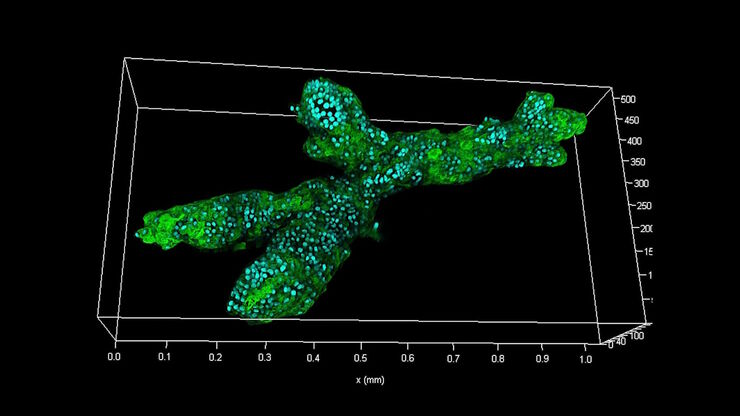
Branched organoid growing in collagen where the Nuclei are labeled blue. To detect the mechanosignaling process, the YAP1 is labeled green.

Examining Developmental Processes In Cancer Organoids

Interview: Prof. Bausch and Dr. Pastucha, Technical University of Munich, discuss using microscopy to study development of organoids, stem cells, and other relevant disease models for biomedical…
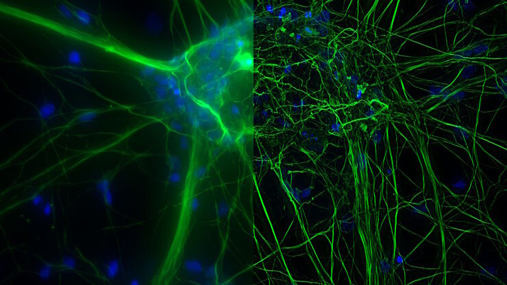
Mouse cortical neurons. Transgenic GFP (green). Image courtesy of Prof. Hui Guo, School of Life Sciences, Central South University, China

How Microscopy Helps the Study of Mechanoceptive and Synaptic Pathways

In this podcast, Dr Langenhan explains how microscopy helps his team to study mechanoceptive and synaptic pathways, their challenges, and how they overcome them.
Advanced technologies support neurosurgical teaching. Image courtesy of Dr. Florian Bernard.

3D, AR & VR for Teaching in Neurosurgery

Discover the evolution of neurosurgical teaching and how 3D, Augmented Reality and Virtual Reality can help better learn anatomy and acquire surgical skills.
THY1-EGFP labeled neurons in mouse brain processed using the PEGASOS 2 tissue clearing method, imaged on a Leica confocal microscope. Neurons were traced using Aivia’s 3D Neuron Analysis – FL recipe. Image credit: Hu Zhao, Chinese Institute for Brain Research.

Unlocking Insights in Complex and Dense Neuron Images Guided by AI

The latest advancement in Aivia AI image analysis software provides improved soma detection, additional flexibility in neuron tracing, 3D relational measurement including Sholl analysis and more.
Microscopy for neuroscience research

What are the Challenges in Neuroscience Microscopy?

eBook outlining the visualization of the nervous system using different types of microscopy techniques and methods to address questions in neuroscience.
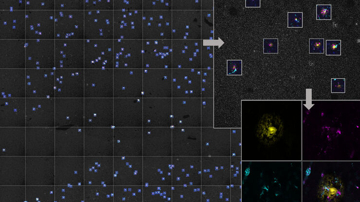
AI-based workflow for fast rare event detection in living biological samples using Autonomous Microscopy powered by Aivia

AI Microscopy Enables the Efficient Detection of Rare Events

Localization and selective imaging of rare events is key for the investigation of many processes in biological samples. Yet, due to time constraints and complexity, some experiments are not feasible…
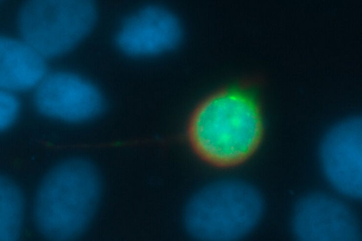
Fluorescence microscope image of a life-science specimen

An Introduction to Fluorescence

This article gives an introduction to fluorescence and photoluminescence, which includes phosphorescence, explains the basic theory behind them, and how fluorescence is used for microscopy.

How to Prepare and Analyse Battery Samples with Electron Microscopy

This workshop covers the sample preparation process for lithium and novel battery sample analysis, as well as other semiconductor samples requiring high-resolution cross-section imaging.
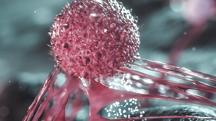
Cancer cells

The Role of Iron Metabolism in Cancer Progression

Iron metabolism plays a role in cancer development and progression, and modulates the immune response. Understanding how iron influences cancer and the immune system can aid the development of new…
Pancreatic Ductal Adenocarcinoma with 5 biomarkers shown – SMA, PanCK PCK26, PanCK AE1, Vimentin, and Glut1.

Dig Deeper Into the Complexities of Pancreatic Cancer with Multiplex Imaging

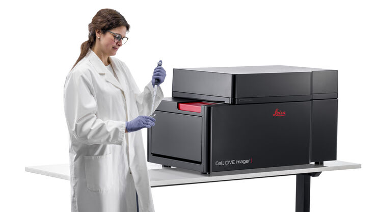
Cell DIVE is an iterative staining workflow for multiplexed imaging that unveils biological pathways to dig deeper into the complexities of pancreatic cancer.
Cell DIVE Multiplex Imaging Solution

Complex Made Simple: Antibodies in Multiplexed Imaging

Build panels, plan studies, and get the most from precious reagents using this antibody multiplexing guide from Leica Microsystems
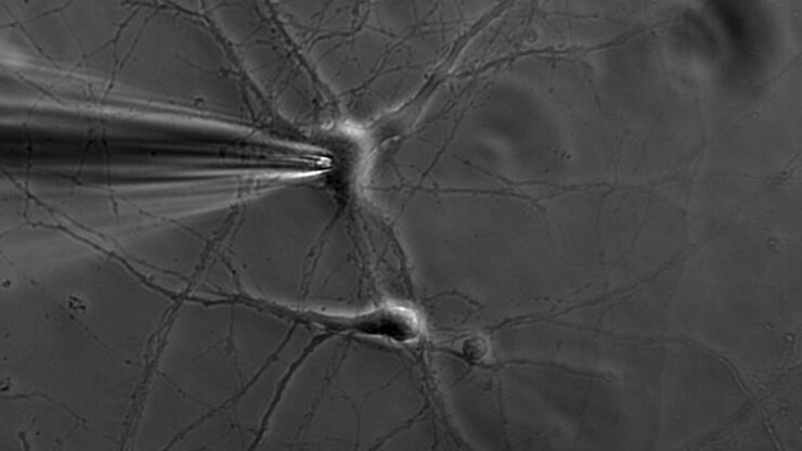
Patch pipette touching a murine hippocampal neuron. Image courtesy of A. Aguado, Ruhr University Bochum, Germany.

What is the Patch-Clamp Technique?

This article gives an introduction to the patch-clamp technique and how it is used to study the physiology of ion channels for neuroscience and other life-science fields.
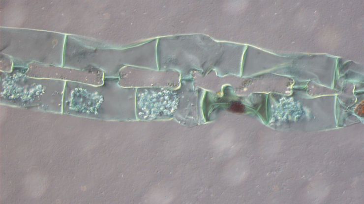
Spirogyra algae (Conjugation), Transmitted Light Differential Interference Contrast.

Differential Interference Contrast (DIC) Microscopy

This article demonstrates how differential interference contrast (DIC) can be actually better than brightfield illumination when using microscopy to image unstained biological specimens.
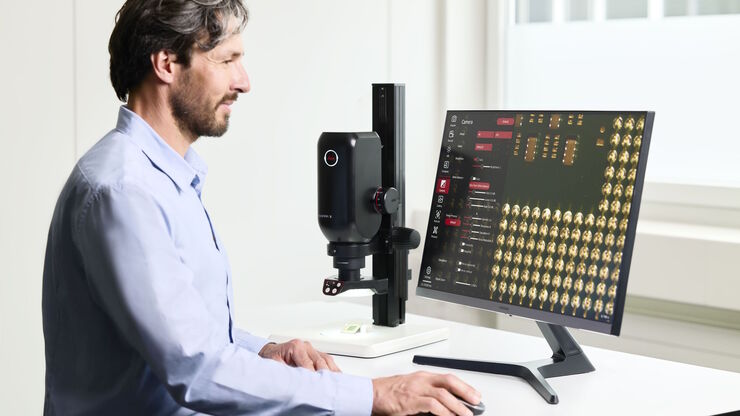
The Emspira 3 digital microscope offers what users need for comprehensive visual inspection, including comparison, measurement, and documentation sharing.

Digital Inspection Microscope for Industrial Applications

Factors users should consider before choosing a digital inspection microscope for industrial applications, including quality control (QC), failure analysis (FA), and R&D, are described in this…

Going Beyond Deconvolution

Widefield fluorescence microscopy is often used to visualize structures in life science specimens and obtain useful information. With the use of fluorescent proteins or dyes, discrete specimen…
Inspection microscope image of a printed circuit board (PCB) taken with a ring light (RL) and near vertical illumination (NVI).

Microscope Illumination for Industrial Applications

Inspection microscope users can obtain information from this article which helps them choose the optimal microscope illumination or lighting system for inspection of parts or components.
Image of MDCK (Madin-Darby canine kidney) cells taken with phase contrast.

Phase Contrast and Microscopy

This article explains phase contrast, an optical microscopy technique, which reveals fine details of unstained, transparent specimens that are difficult to see with common brightfield illumination.

Immersion Objectives

How an immersion objective, which has a liquid medium between it and the specimen being observed, helps increase the numerical aperture and microscope resolution is explained in this article.
Phase-contrast image of a MDCK-cell culture and its respective confluency measured by the Mateo TL microscope.

How to Determine Cell Confluency with a Digital Microscope

This article shows how to measure cell confluency in an easy and consistent way with Mateo TL, increasing confidence in downstream experiments.
Microscope image of cultured cells at the bottom of a dish.

How to do a Proper Cell Culture Quick Check

In order to successfully work with mammalian cell lines, they must be grown under controlled conditions and require their own specific growth medium. In addition, to guarantee consistency their growth…
Scroll to top