
Science Lab
Science Lab
Das Wissensportal von Leica Microsystems bietet Ihnen Wissens- und Lehrmaterial zu den Themen der Mikroskopie. Die Inhalte sind so konzipiert, dass sie Einsteiger, erfahrene Praktiker und Wissenschaftler gleichermaßen bei ihrem alltäglichen Vorgehen und Experimenten unterstützen. Entdecken Sie interaktive Tutorials und Anwendungsberichte, erfahren Sie mehr über die Grundlagen der Mikroskopie und High-End-Technologien - werden Sie Teil der Science Lab Community und teilen Sie Ihr Wissen!
Loading...

The Power HyD Detector Family
Powerful photon counting detectors on the STELLARIS confocal platform provide improved photon counting, ultra-sensitive imaging and more color options in the NIR spectrum.
Loading...
How to Uncover Hidden Dimensions in Research with Lifetime Imaging
Learn how fluorescence lifetime imaging adds information depth to your confocal experiments and reveals novel insights that are difficult or impossible to discover using conventional intensity-based…
Loading...
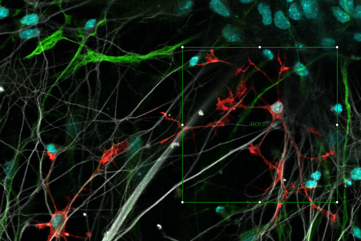
 from the desired fluorescent signal from the cell membrane (green).")
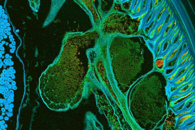
Learn how to Remove Autofluorescence from your Confocal Images
Autofluorescence can significantly reduce what you can see in a confocal experiment. This article explores causes of autofluorescence as well as different ways to remove it, from simple media fixes to…
Loading...

Zebrafish Brain - Whole Organ Imaging at High Resolution
Structural information is key when one seeks to understand complex biological systems, and one of the most complex biological structures is the vertebrate central nervous system. To image a complete…
Loading...
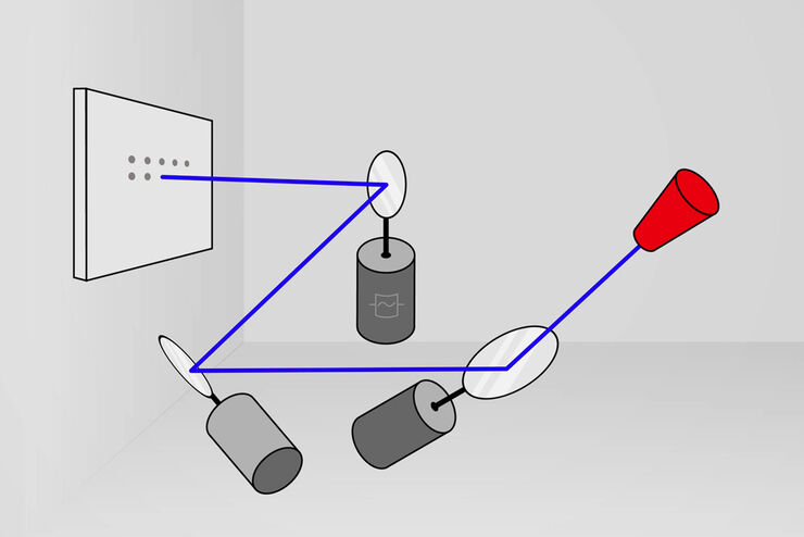
What is a Resonant Scanner?
A resonant scanner is a type of galvanometric mirror scanner that allows fast image acquisition with single-point scanning microscopes (true confocal and multiphoton laser scanning). High acquisition…
Loading...
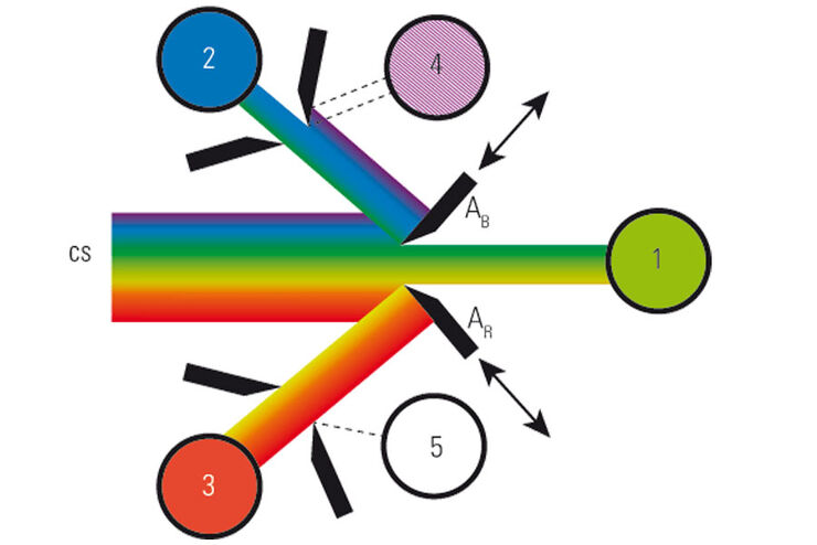
What is a Spectral Detector (SP Detector)?
The SP detector from Leica Microsystems denotes a compound detection unit for point scanning microscopes, in particular confocal microscopes. The SP detector splits light into up to 5 spectral bands.…
Loading...

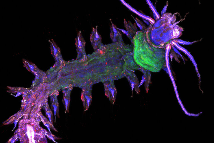
Improve 3D Cell Biology Workflow with Light Sheet Microscopy
Understanding the sub-cellular mechanisms in carcinogenesis is of crucial importance for cancer treatment. Popular cellular models comprise cancer cells grown as monolayers. But this approach…
Loading...
What is a Field-of-View Scanner?
A field-of-view scanner is an assembly of galvanometric scanning mirrors used in single-point confocal microscopes that offer the correct optical recording of large field sizes. The field-of-view…
Loading...
Resolved Field Number (RFN)
The field number (FN) for optical microscopes indicates the field of view (FOV). It corresponds to the area in the intermediate image that is observable through the eyepieces. Although, we cannot…