
Science Lab
Science Lab
Das Wissensportal von Leica Microsystems bietet Ihnen Wissens- und Lehrmaterial zu den Themen der Mikroskopie. Die Inhalte sind so konzipiert, dass sie Einsteiger, erfahrene Praktiker und Wissenschaftler gleichermaßen bei ihrem alltäglichen Vorgehen und Experimenten unterstützen. Entdecken Sie interaktive Tutorials und Anwendungsberichte, erfahren Sie mehr über die Grundlagen der Mikroskopie und High-End-Technologien - werden Sie Teil der Science Lab Community und teilen Sie Ihr Wissen!
Loading...
Spectroscopic Evaluation of Red Blood Cells
Hemoglobinopathies are a major healthcare problem. This study presents a possible diagnostic tool for thalassemia which is based on confocal spectroscopy. This approach exploits spectral detection and…
Loading...
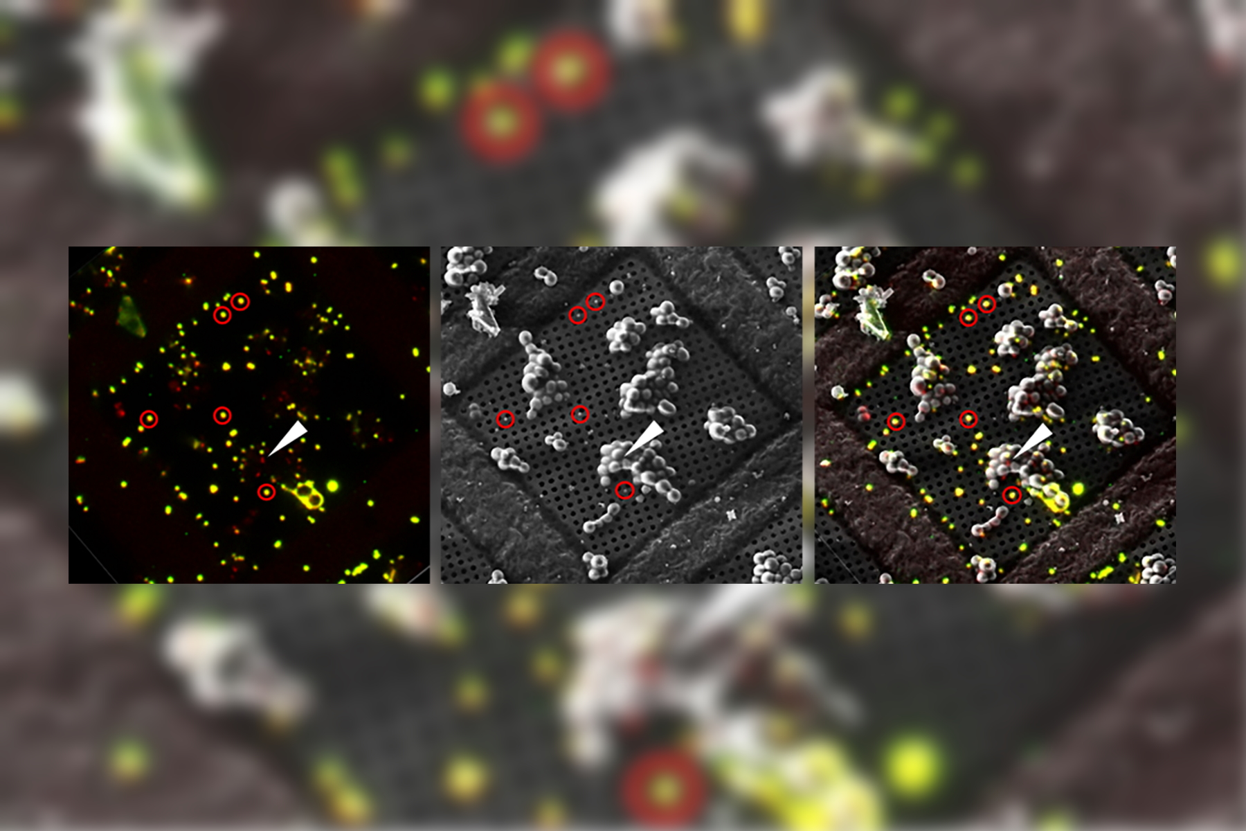
The Cryo-CLEM Journey
This article describes the Cryo-CLEM technology and the benefits it can provide for scientists. Additionally, some scientific publications are highlighted.
Recent developments in cryo electron…
Loading...
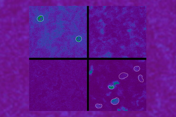
DNA Replication in Cancer Cells
DNA synthesis can be impeded by collisions between the DNA replication machinery and co-transcriptional R-loops leading to a major source of genomic instability in cancer cells. In this paper we…
Loading...
Fluorescence Lifetime-based Imaging Gallery
Confocal microscopy relies on the effective excitation of fluorescence probes and the efficient collection of photons emitted from the fluorescence process. One aspect of fluorescence is the emission…
Loading...
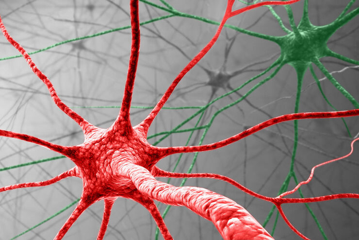
Visualizing Protein Degradation and Aggregation in the Living Cell
Our guest speaker, Prof Dr Eric Reits, presents his work on neurodegenerative disorders. Reits’ group are experts on the subject of Huntington’s disease and work towards identifying leads for…
Loading...
Life Beyond the Pixels: Deep Learning Methods for Single Cell Analysis
Our guest speaker Prof Dr Peter Horvath presents his work on single cell-based large-scale microscopy experiments. This novel targeting approach includes the use of machine learning models and…
Loading...
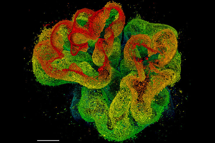
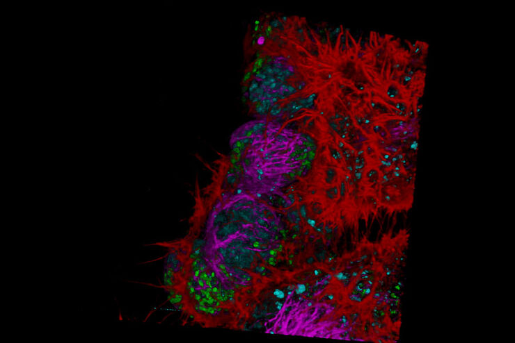
Tissue Image Gallery
Visual analysis of animal and human tissues is critical to understand complex diseases such as cancer or neurodegeneration. From basic immunohistochemistry to intravital imaging, confocal microscopy…
Loading...
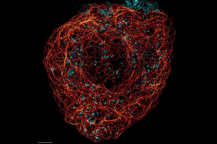
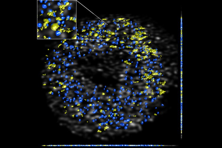
Super-Resolution Microscopy Image Gallery
Due to the diffraction limit of light, traditional confocal microscopy cannot resolve structures below ~240 nm. Super-resolution microscopy techniques, such as STED, PALM or STORM or some…
Loading...

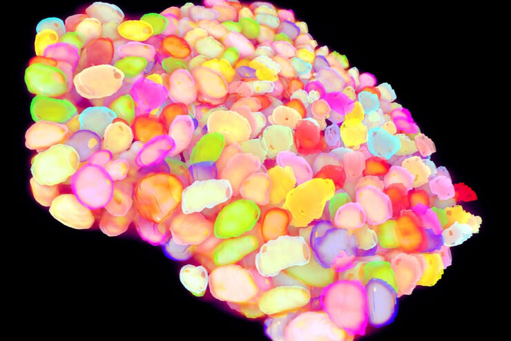
Live Cell Imaging Gallery
Live cell microscopy techniques are fundamental to get a better understanding of cellular and molecular function. Today, widefield microscopy is the most common technique used to visualize cell…