
Science Lab
Science Lab
Das Wissensportal von Leica Microsystems bietet Ihnen Wissens- und Lehrmaterial zu den Themen der Mikroskopie. Die Inhalte sind so konzipiert, dass sie Einsteiger, erfahrene Praktiker und Wissenschaftler gleichermaßen bei ihrem alltäglichen Vorgehen und Experimenten unterstützen. Entdecken Sie interaktive Tutorials und Anwendungsberichte, erfahren Sie mehr über die Grundlagen der Mikroskopie und High-End-Technologien - werden Sie Teil der Science Lab Community und teilen Sie Ihr Wissen!
Loading...

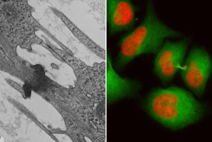
Precise 3D Targeting for EM Imaging - Access What Matters
Find out how the seamless cryo-electron tomography workflow Coral Cryo uses confocal super resolution to target your structure of interest more precisely.
Loading...
![[Translate to German:] SEM image of the full Li-NMC electrode sample, showing the two porous layers and the metal film at the center of the structure.](/fileadmin/_processed_/2/f/csm_Cross_Section_Ion_Beam_Milling_of_Battery_Components_teaser_5295ac1cbc.jpg "[Translate to German:] SEM image of the full Li-NMC electrode sample, showing the two porous layers and the metal film at the center of the structure.")
Querschnitt-Ionenstrahlfräsen von Batteriekomponenten
Für ein umfassendes Verständnis von Lithiumbatteriesystemen ist eine qualitativ hochwertige Oberflächenpräparation erforderlich, um die innere Struktur und Morphologie zu untersuchen. Aufgrund der…
Loading...
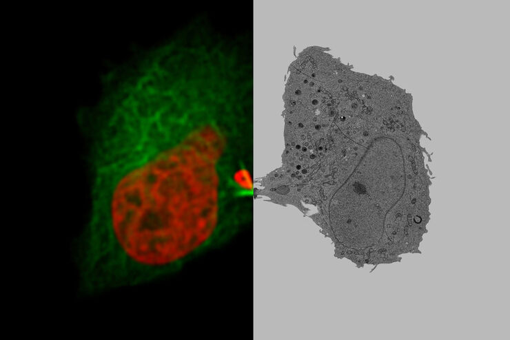
How to Successfully Implement Coral Life
The live-cell CLEM workflow allows you to capture dynamic information related to a relevant biological process as it happens and put these observations into their ultrastructural context. The Leica…
Loading...

How to Improve Live Cell Imaging with Coral Life
For live-cell CLEM applications, light microscopy imaging is a critical step for identifying the right cell in the right state at the right time. In this article, Leica experts share their insights on…
Loading...
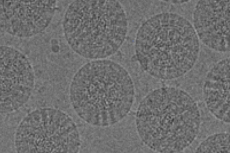
The Cryo-CLEM Journey
This article describes the Cryo-CLEM technology and the benefits it can provide for scientists. Additionally, some scientific publications are highlighted.
Recent developments in cryo electron…
Loading...

How to Keep Your Samples Under Physiological Conditions
The Coral Life workflow combines dynamic data with the best possible sample fixation by high pressure freezing. However, good sample preservation won’t help if your cells are stressed by temperature…
Loading...
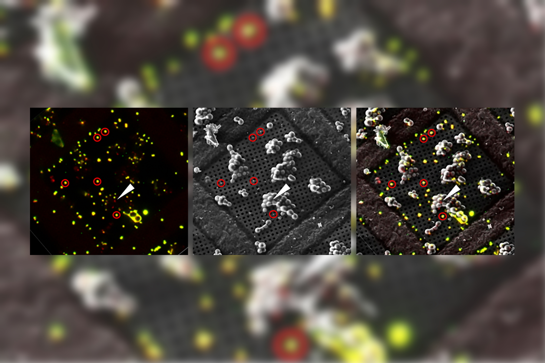
Putting Dynamic Live Cell Data into the Ultrastructural Context
With workflow Coral Life, searching for a needle in the haystack is a thing of the past. Take advantage of correlative light and electron microscopy to identify directly the right cell at the right…
Loading...

Download EM Workflow Solutions Booklet
This publication is a compilation of appropriate workflows for the most frequently used sample preparation methods, like Correlative Methodologies, Optogenetics & Electro-Physiology, Surface Analysis,…
Loading...
Exploring the Structure and Life Cycle of Viruses
The SARS-CoV-2 outbreak started in late December 2019 and has since reached a global pandemic, leading to a worldwide battle against COVID-19. The ever-evolving electron microscopy methods offer a…