
Science Lab
Science Lab
Das Wissensportal von Leica Microsystems bietet Ihnen Wissens- und Lehrmaterial zu den Themen der Mikroskopie. Die Inhalte sind so konzipiert, dass sie Einsteiger, erfahrene Praktiker und Wissenschaftler gleichermaßen bei ihrem alltäglichen Vorgehen und Experimenten unterstützen. Entdecken Sie interaktive Tutorials und Anwendungsberichte, erfahren Sie mehr über die Grundlagen der Mikroskopie und High-End-Technologien - werden Sie Teil der Science Lab Community und teilen Sie Ihr Wissen!
Loading...
, actin network (ATTO 647N), and nuclear pore basket (CF 680R).")
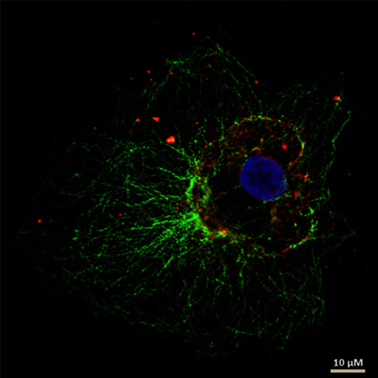
The Guide to STED Sample Preparation
This guide is intended to help users optimize sample preparation for stimulated emission depletion (STED) nanoscopy, specifically when using the STED microscope from Leica Microsystems. It gives an…
Loading...
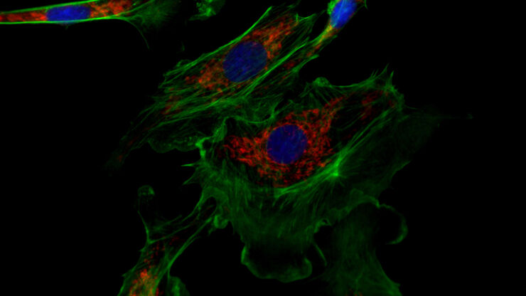
Erforschung der Virusreplikaton mit Fluoreszenzmikroskopie
Viren können mit Hilfe verschiedener Mikroskopietechniken untersucht werden. Je nach Vergrößerung und Auflösung des Mikroskops kann die Beobachtung auf Gewebe-, Zell- oder Virionenebene erfolgen.
Loading...
![[Translate to German:] Cell counts for each biomarker were divided by total number of cells to give a percentage of biomarker positive cells out of total cells for each biomarker.](/fileadmin/_processed_/5/3/csm_Cell_counts_for_biomarkers_with_Cell_DIVE_ee648b397a.jpg "[Translate to German:] Cell counts for each biomarker were divided by total number of cells to give a percentage of biomarker positive cells out of total cells for each biomarker.")
Methoden zur Verbesserung der Reproduzierbarkeit in der räumlichen Biologieforschung
Durch den Einsatz von Automatisierung, hochwertigen Antikörpern und einem bewährten Multiplex-Imaging-Workflow ist Cell DIVE in der Lage, reproduzierbare Ergebnisse zu liefern. In diesem Poster…
Loading...
 tissue on a single slide.")
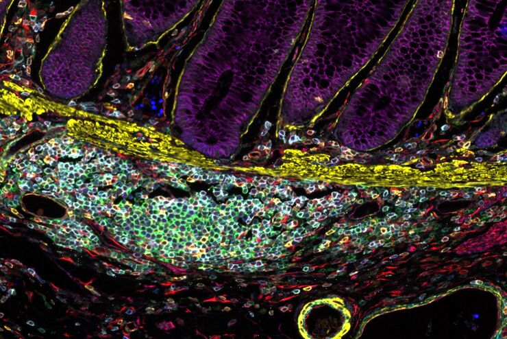
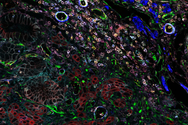
Characterizing tumor environment to reveal insights and spatial resolution
Antibodies from Cell Signaling Technology are validated for use with the Cell DIVE multiplexing workflow and used to probe cell lineages in the tumor microenvironment
Loading...

Multiplexed Imaging Types, Benefits and Applications
Multiplexed imaging is an emerging and exciting way to extract information from human tissue samples by visualizing many more biomarkers than traditional microscopy. By observing many biomarkers…
Loading...
Wie Sie Gewebeproben für die Immunfluoreszenz-Mikroskopie vorbereiten
Immunfluoreszenz (IF) ist eine leistungsfähige Methode zur Visualisierung intrazellulärer Prozesse, Bedingungen und Strukturen. IF-Präparate können mit verschiedenen Mikroskopietechniken (z. B. CLSM,…
Loading...
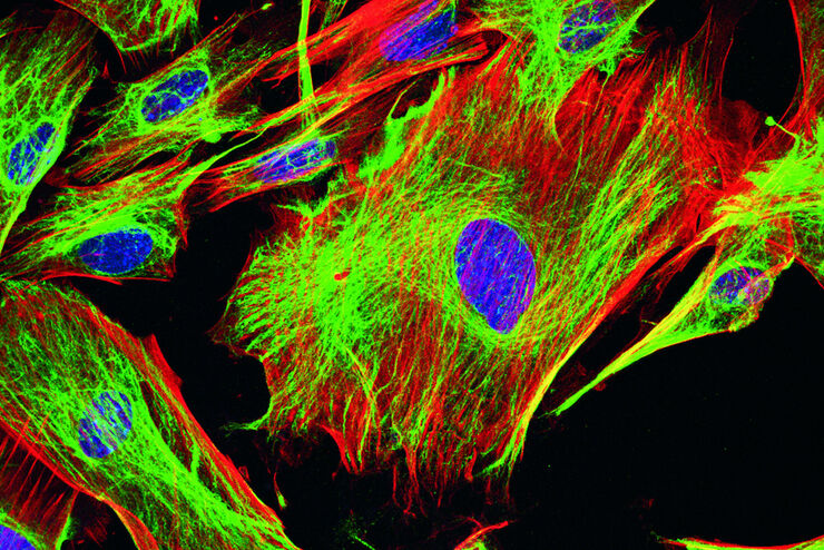
Fluorescent Dyes
A basic principle in fluorescence microscopy is the highly specific visualization of cellular components with the help of a fluorescent agent. This can be a fluorescent protein – for example GFP –…
Loading...
Designing your Research Study with Multiplexed IF Imaging
Multiplexed tissue analysis is a powerful technique that allows comparisons of cell-type locations and cell-type interactions within a single fixed tissue sample. It is common for researchers to ask…
Loading...
Be Confident in your Results with Cell DIVE Validated Antibodies
The Cell DIVE System includes a carefully curated list of hundreds of commercially available antibodies validated to offer optimal specificity and sensitivity in multiplexed imaging. That validation…