
Science Lab
Science Lab
Das Wissensportal von Leica Microsystems bietet Ihnen Wissens- und Lehrmaterial zu den Themen der Mikroskopie. Die Inhalte sind so konzipiert, dass sie Einsteiger, erfahrene Praktiker und Wissenschaftler gleichermaßen bei ihrem alltäglichen Vorgehen und Experimenten unterstützen. Entdecken Sie interaktive Tutorials und Anwendungsberichte, erfahren Sie mehr über die Grundlagen der Mikroskopie und High-End-Technologien - werden Sie Teil der Science Lab Community und teilen Sie Ihr Wissen!
Loading...
A New Method for Convenient and Efficient Multicolor Imaging
The technique combining hyperspectral unmixing and phasor analysis was developed to simplify the process of getting images from a sample labeled with multiple fluorophores. This aggregate method…
Loading...
Considerations for Multiplex Live Cell Imaging
Simultaneous multicolor imaging for successful experiments: Live-cell imaging experiments are key to understand dynamic processes. They allow us to visually record cells in their living state, without…
Loading...
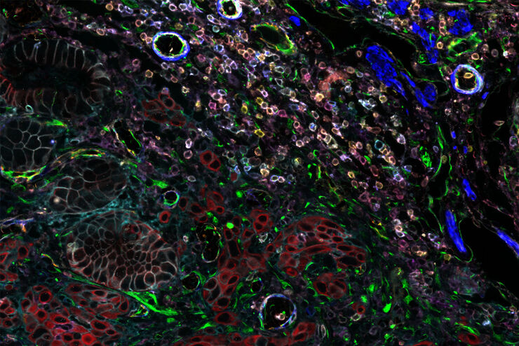
Designing your Research Study with Multiplexed IF Imaging
Multiplexed tissue analysis is a powerful technique that allows comparisons of cell-type locations and cell-type interactions within a single fixed tissue sample. It is common for researchers to ask…
Loading...
How to Successfully Perform Live-cell CLEM
The Leica Nano workflow provides a streamlined live-cell CLEM solution for getting insight bout structural changes of cellular components over time. Besides the technical handling described in the…
Loading...
Be Confident in your Results with Cell DIVE Validated Antibodies
The Cell DIVE System includes a carefully curated list of hundreds of commercially available antibodies validated to offer optimal specificity and sensitivity in multiplexed imaging. That validation…
Loading...
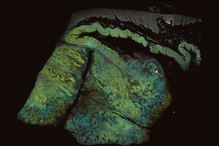
Overcoming Multiplexed Imaging Roadblocks
Whether a single slide or a batch of slides processed on multiple Cell DIVE imagers, every step carried out by the Cell DIVE Acquisition Software eliminates distortions and artifacts and seamlessly…
Loading...

An Introduction to Computational Clearing
Many software packages include background subtraction algorithms to enhance the contrast of features in the image by reducing background noise. The most common methods used to remove background noise…