
Science Lab
Science Lab
Das Wissensportal von Leica Microsystems bietet Ihnen Wissens- und Lehrmaterial zu den Themen der Mikroskopie. Die Inhalte sind so konzipiert, dass sie Einsteiger, erfahrene Praktiker und Wissenschaftler gleichermaßen bei ihrem alltäglichen Vorgehen und Experimenten unterstützen. Entdecken Sie interaktive Tutorials und Anwendungsberichte, erfahren Sie mehr über die Grundlagen der Mikroskopie und High-End-Technologien - werden Sie Teil der Science Lab Community und teilen Sie Ihr Wissen!
Loading...
![[Translate to German:] Co-detection of 10 extracellular matrix proteins and 3 topographical tissue landmarks by multiplex immunostaining within a single high-grade fibrous hotspot from a human hepatocellular carcinoma [Translate to German:] Co-detection of 10 extracellular matrix proteins and 3 topographical tissue landmarks by multiplex immunostaining within a single high-grade fibrous hotspot from a human hepatocellular carcinoma](/fileadmin/_processed_/0/8/csm_Single_high-grade_fibrous_hotspot_from_a_human_hepatocellular_carcinoma_e5541282bd.jpg)
In-situ-Identifizierung von Krebsstammzellnischen im Leberzellkarzinom
Die Multiplex-Bildgebung spielt eine entscheidende Rolle bei der Krebsforschung, da sie es Wissenschaftlern ermöglicht, tief in die komplexe Mikroumgebung von Tumoren einzudringen und einzigartige…
Loading...
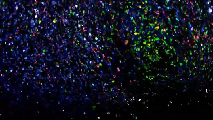
Potential of Multiplex Confocal Imaging for Cancer Research and Immunology
Explore the new frontiers of multi-color fluorescent imaging: from image acquisition to analysis
Loading...
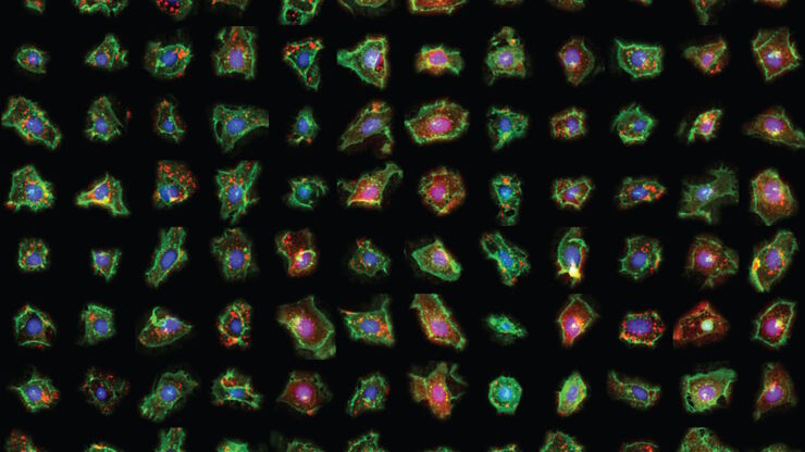
Exploring Subcellular Spatial Phenotypes with SPARCS
Discover spatially resolved CRISPR screening (SPARCS), a platform for microscopy-based genetic screening for spatial subcellular phenotypes at the human genome scale.
Loading...
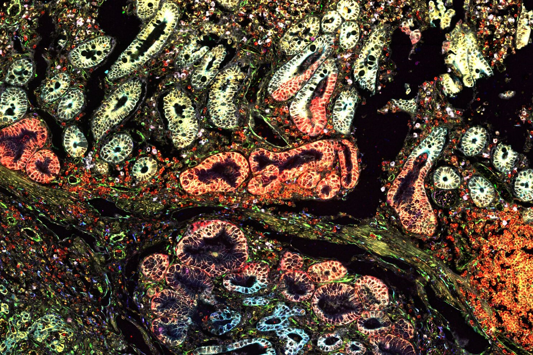
Multiplexing with Luke Gammon: Advance your Spatial Biology Research
Learn how multiplexing imaging and spatial biology can help researchers better understand complex biological systems. In this interview, Dr. Gammon and Dr. Pointu of Leica Microsystems discuss pain…
Loading...
![[Translate to German:] Multi-tissue array with 4 markers shown including DAPI, NaKATPase, PanCk, and Vimentin. [Translate to German:] Multi-tissue array with 4 markers shown including DAPI, NaKATPase, PanCk, and Vimentin.](/fileadmin/_processed_/8/b/csm_Multi-tissue_array_with_4_markers_93819abf05.jpg)
Räumliche Biologie: Erwägung neuer Wege
Räumliche Biologie: Forschung zu Anordnung und Interaktion von Molekülen, Zellen und Geweben in ihrem nativen räumlichen Kontext
Loading...
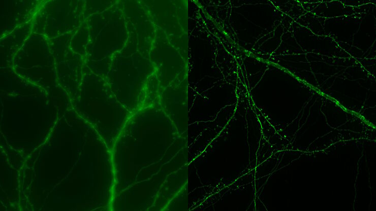
How Microscopy Helps the Study of Mechanoceptive and Synaptic Pathways
In this podcast, Dr Langenhan explains how microscopy helps his team to study mechanoceptive and synaptic pathways, their challenges, and how they overcome them.
Loading...
![[Translate to German:] Cell counts for each biomarker were divided by total number of cells to give a percentage of biomarker positive cells out of total cells for each biomarker. [Translate to German:] Cell counts for each biomarker were divided by total number of cells to give a percentage of biomarker positive cells out of total cells for each biomarker.](/fileadmin/_processed_/5/3/csm_Cell_counts_for_biomarkers_with_Cell_DIVE_ee648b397a.jpg)
Methoden zur Verbesserung der Reproduzierbarkeit in der räumlichen Biologieforschung
Durch den Einsatz von Automatisierung, hochwertigen Antikörpern und einem bewährten Multiplex-Imaging-Workflow ist Cell DIVE in der Lage, reproduzierbare Ergebnisse zu liefern. In diesem Poster…
Loading...
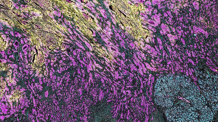
Characterizing tumor environment to reveal insights and spatial resolution
Antibodies from Cell Signaling Technology are validated for use with the Cell DIVE multiplexing workflow and used to probe cell lineages in the tumor microenvironment
Loading...

How is Microscopy Used in Spatial Biology? A Microscopy Guide
Different spatial biology methods in microscopy, such as multiplex imaging, are helping to better understand tissue landscapes. Learn more in this microscopy guide.