
Science Lab
Science Lab
Das Wissensportal von Leica Microsystems bietet Ihnen Wissens- und Lehrmaterial zu den Themen der Mikroskopie. Die Inhalte sind so konzipiert, dass sie Einsteiger, erfahrene Praktiker und Wissenschaftler gleichermaßen bei ihrem alltäglichen Vorgehen und Experimenten unterstützen. Entdecken Sie interaktive Tutorials und Anwendungsberichte, erfahren Sie mehr über die Grundlagen der Mikroskopie und High-End-Technologien - werden Sie Teil der Science Lab Community und teilen Sie Ihr Wissen!
Loading...
Confocal Imaging of Immune Cells in Tissue Samples
In this webinar, you will discover how to perform 10-color acquisition using a confocal microscope. The challenges of imaged-based approaches to identify skin immune cells. A new pipeline to assess…
Loading...
FluoSync - a Fast & Gentle Method for Unmixing Multicolour Images
In this white paper, we focus on a fast and reliable method for obtaining high-quality multiplex images in fluorescence microscopy. FluoSync combines an existing method for hybrid unmixing with…
Loading...

Multiplexing through Spectral Separation of 11 Colors
Fluorescence microscopy is a fundamental tool for life science research that has evolved and matured together with the development of multicolor labeling strategies in cells tissues and model…
Loading...

Multiplexed Imaging Types, Benefits and Applications
Multiplexed imaging is an emerging and exciting way to extract information from human tissue samples by visualizing many more biomarkers than traditional microscopy. By observing many biomarkers…
Loading...

Multiplexed Imaging Glossary
Human tissues contain a many different biomarkers which can be explored to reveal the workings of biological pathways and interrogate disease states. Recent technological advances have made it…
Loading...

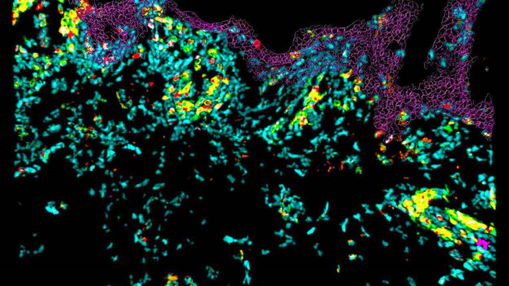
Tumor MHC Expression and Intralesional IL2 Response in Melanoma
Genomics profiling and Cell DIVE multiplex imaging allows researchers to understand the immune cell phenotypes that most strongly predict response to IL2 immunotherapy in melanoma patients suffering…
Loading...

Create New Options for Live Cell Imaging
The use of state-of-the-art AI systems is pushing image analysis into a new generation. Challenges like the conflict between imaging power and sample integrity are being overcome with THUNDER’s…
Loading...

How to Target Fluorescent Structures in 3D for Cryo-FIB Milling
This article describes the major steps of the cryo-electron tomography workflow including super-resolution cryo-confocal microscopy. We describe how subcellular structures can be precisely located in…
Loading...
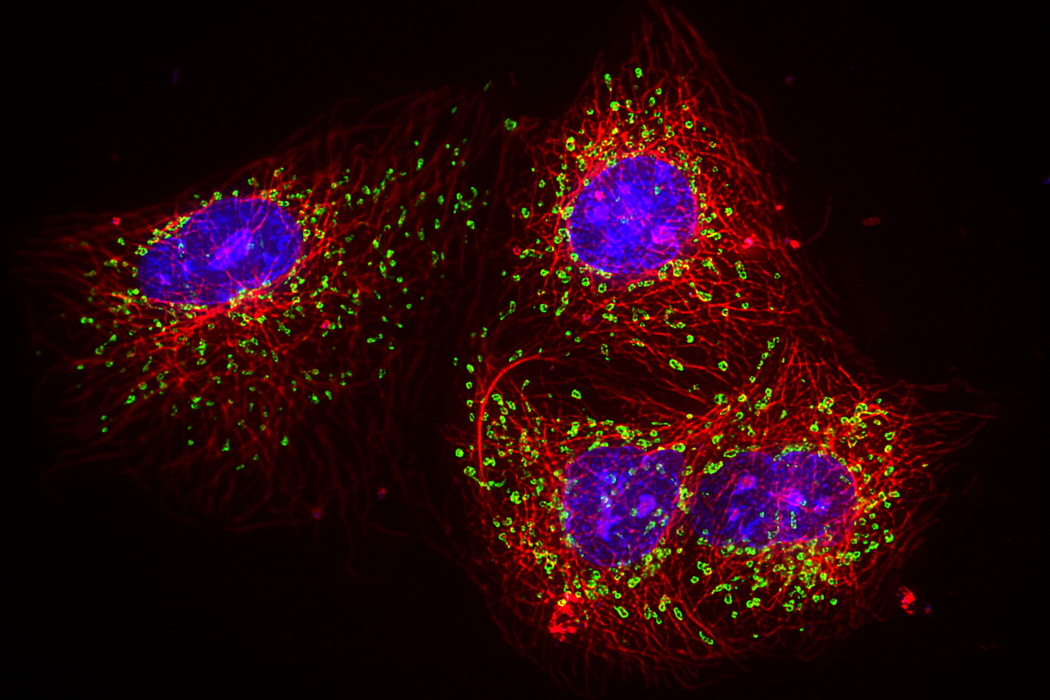
, Astrocytes (GFAP, red), Nuclei (DAPI, blue).")
Multicolor Microscopy: The Importance of Multiplexing
The term multiplexing refers to the use of multiple fluorescent dyes to examine various elements within a sample. Multiplexing allows related components and processes to be observed in parallel,…