
Science Lab
Science Lab
Bienvenido al portal de conocimiento de Leica Microsystems. Aquí encontrará investigación científica y material didáctico sobre el tema de la microscopía. El portal ayuda a principiantes, profesionales experimentados y científicos por igual en su trabajo diario y en sus experimentos. Explore tutoriales interactivos y notas de aplicación, descubra los fundamentos de la microscopía, así como las tecnologías de gama alta. Forme parte de la comunidad Science Lab y comparta sus conocimientos.
Filter articles
Tags
Story Type
Products
Loading...
Workflow Solutions for Sample Preparation Methods for Material Science
This brochure presents and explains appropriate workflow solutions for the most frequently required sample preparation methods for material science samples.
Loading...

Automatic Alignment of Sample and Knife for High Sectioning Quality
Automatic alignment of sample and knife on the ultramicrotome UC Enuity, enabling even untrained users to create ultrathin sections with reduced risk of losing precious sections.
Loading...

High Quality Sectioning in Ultramicrotomy
Discover the significance of achieving high-quality uniform sections with ultramicrotomy for precise imaging in electron microscopy.
Loading...

Quality Control via Cross Sections of PCBs, PCBAs, ICs, and Batteries
Why cross sections of printed circuit boards (PCBs) and assemblies (PCBAs), integrated circuits (ICs), and battery components are useful for quality control (QC), failure analysis (FA), and research…
Loading...

Studying Virus Replication with Fluorescence Microscopy
The results from research on SARS-CoV-2 virus replication kinetics, adaption capabilities, and cytopathology in Vero E6 cells, done with the help of fluorescence microscopy, are described in this…
Loading...

Five Inverted-Microscope Advantages for Industrial Applications
With inverted microscopes, you look at samples from below since their optics are placed under the sample, with upright microscopes you look at samples from above. Traditionally, inverted microscopes…
Loading...
 stained with osmium tetroxide (OsO4), sectioned with a DIATOME diamond knife at room temperature, and then imaged with HAADF TEM.")
Ultramicrotomy Techniques for Materials Sectioning
Learn about ultramicrotomy for materials sectioning when investigating polymers and brittle materials with transmission (TEM) or scanning electron microscopy (SEM) or atomic force microscopy.
Loading...
 chip cross section acquired at higher magnification showing a region of interest.")
Structural and Chemical Analysis of IC-Chip Cross Sections
This article shows how electronic IC-chip cross sections can be efficiently and reliably prepared and then analyzed, both visually and chemically at the microscale, with the EM TXP and DM6 M LIBS…
Loading...
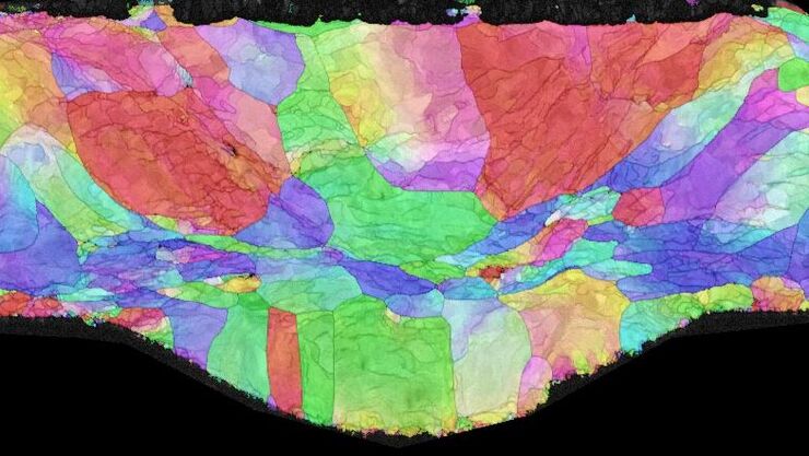
. The grains are highlighted with arbitrary colors.")
High-Quality EBSD Sample Preparation
This article describes a method for EBSD sample preparation of challenging materials. The high-quality samples required for electron backscatter diffraction are prepared with broad ion-beam milling.
