Corporate Communications

Leica Microsystems develops and manufactures microscopes and scientific instruments for the analysis of microstructures and nanostructures.
We offer scientific research and teaching material on the subjects of microscopy. The content is designed to support beginners, experienced practitioners and scientists alike in their everyday work and experiments. Explore interactive tutorials and application notes, discover the basics of microscopy as well as high-end technologies.
Follow us

Image Gallery: THUNDER Imager
To help you answer important scientific questions, THUNDER Imagers eliminate the out-of-focus blur that clouds the view of thick samples when using camera-based fluorescence microscopes. They achieve…

Plastic & Reconstructive Surgery: Why Use a Microscope
Plastic and Reconstructive Surgery procedures can be delicate. Visualization solutions play an essential role, allowing to perform the surgery in the best conditions. And more and more plastic…

Minimally Invasive Spine Surgery: Improving Precision and Accuracy with Microscopes
Spine surgery is extremely delicate and requires extensive training and experience. Innovative visualization technologies can also help achieve better outcomes allowing to see more and have a clearer…

A Guide to Model Organisms in Research
A model organism is a species used by researchers to study specific biological processes. They have similar genetic characteristics to humans and are commonly used in research areas such as genetics,…

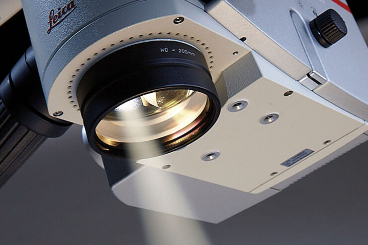
Obtain Maximum Information from your Specimen with LIGHTNING
LIGHTNING is an adaptive process for extraction of information that reveals fine structures and details, otherwise simply not visible, fully automatically. Unlike traditional technologies, that use a…
Advanced Techniques in Cataract and Refractive Surgery
In this webinar Dr. Thompson and Dr. Moshirfar will explain how Leica microscopes aid in procedures such as Centration of Multifocal IOLs and corneal inlays such as Kamra and Lenticular Grafts used in…

A Practical Guide to Virology Research
Leica solutions for imaging and sample preparation help you with the investigation of viral entry and fusion, genome integration, viral replication, assembly, and virus budding.

Surgical Microscopes: Key Factors for OR Nurses
Operating room (OR) nurses are vital to the surgery process. An OR Nurse Manager explains the key surgical microscope features to facilitate their work.

Computational Clearing - Enhance 3D Specimen Imaging
This webinar is designed to clarify crucial specifications that contribute to THUNDER Imagers' transformative visualization of 3D samples and improvements within a researcher's imaging-related…

STELLARIS White Light Lasers
When it comes to choosing fluorescent probes for your multi-color experiments, you shouldn’t have to compromise. Now you can advance beyond conventional excitation sources that limit your fluorophore…
, and trafficking vesicles labeled with CF594 (cyan - Biotium).")
A Guide to Super-Resolution
Find out more about Leica super-resolution microscopy solutions and how they can empower you to visualize in fine detail subcellular structures and dynamics.

Guide to Microscopy in Cancer Research
Cancer is a complex and heterogeneous disease caused by cells deficient in growth regulation. Genetic and epigenetic changes in one or a group of cells disrupt normal function and result in…
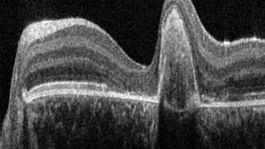
A Guide to OCT
Leica Optical Coherence Tomography (OCT) systems support ophthalmologists, ophthalmic surgeons, and researchers with easy-to-use, high-quality imaging technology.

How to Drape an Overhead Surgical Microscope
The tutorial features the Leica ARveo digital Augmented Reality microscope for complex neurosurgery. The procedure also applies to the Leica M530 OHX, OH6, OH5 and OH4.

How to Drape a Surgical Microscope
Before performing surgical procedures, it is important to drape the surgical microscope to ensure sterile working conditions. At Leica, we are committed to helping you with your surgical practice. In…
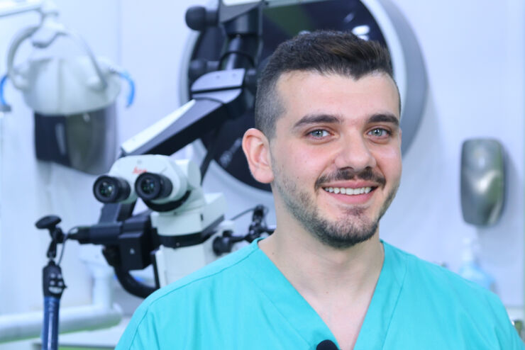
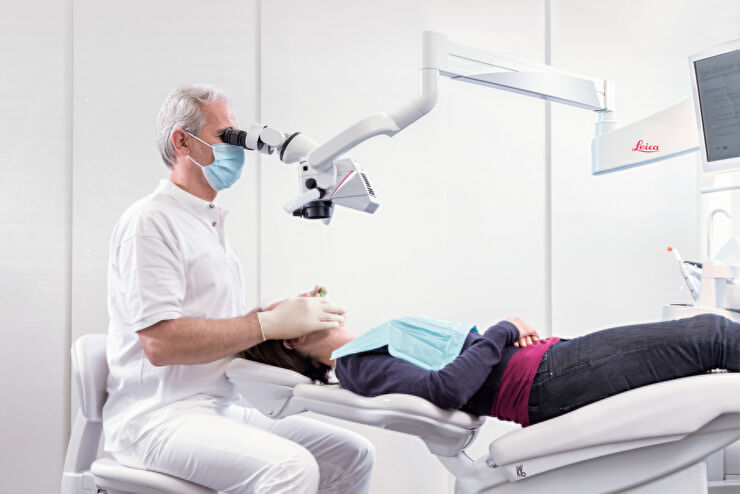
Overcoming Complexities in Microdentistry
Dr. Salam Abu Arqub, from the Smile Engineer Dental Center in Amman, Jordan, has been using Leica dental microscopes for three years for all procedures performed at the clinic. He shared his…

THUNDER Imagers: High Performance, Versatility and Ease-of-Use for your Everyday Imaging Workflows
This webinar will showcase the versatility and performance of THUNDER Imagers in many different life science applications: from counting nuclei in retina sections and RNA molecules in cancer tissue…
Improve Cryo Electron Tomography Workflow
Leica Microsystems and Thermo Fisher Scientific have collaborated to create a fully integrated cryo-tomography workflow that responds to these research needs: Reveal cellular mechanisms at…

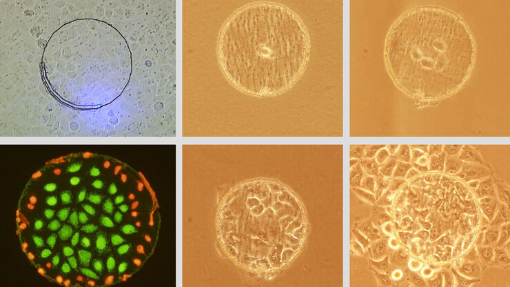
Improve 3D Cell Biology Workflow with Light Sheet Microscopy
Understanding the sub-cellular mechanisms in carcinogenesis is of crucial importance for cancer treatment. Popular cellular models comprise cancer cells grown as monolayers. But this approach…
Live Cell Isolation by Laser Microdissection
Laser microdissection is a tool for the isolation of homogenous cell populations from their native niches in tissues to downstream molecular assays. Beside its routine use for fixed tissue sections,…
Successful Endodontic Treatment with Dental Operating Microscopes
In endodontics, accurate treatment is not only dependent on the technical skills and knowledge of the dentist, but also on clear, detailed visualization of the surgical field. As the outcome of an…
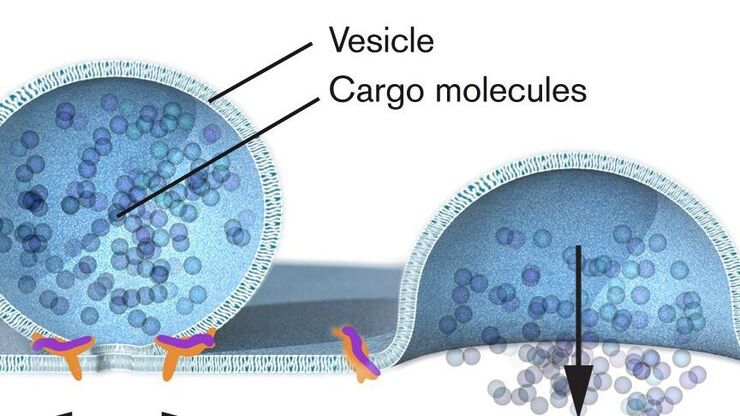
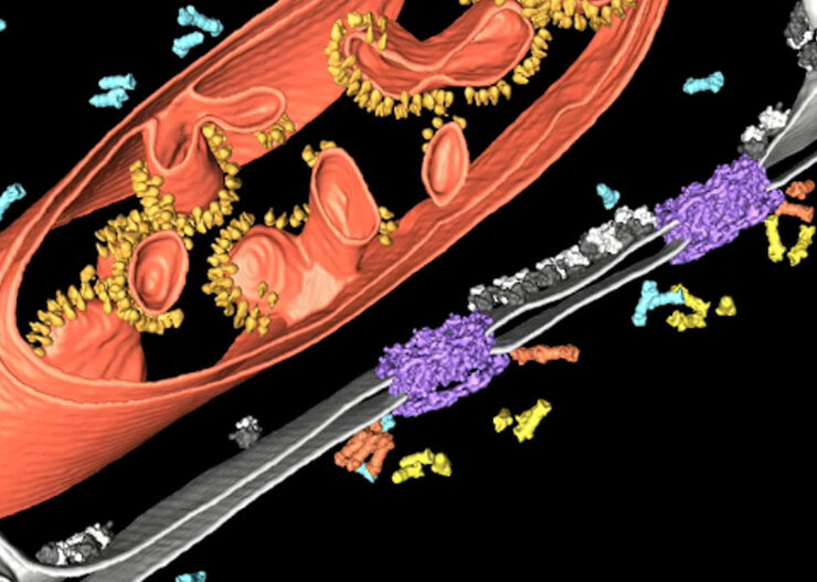
Nobel Prize 2013 in Physiology or Medicine for Discoveries of the Machinery Regulating Vesicle Traffic
On October 7th 2013, The Nobel Assembly at Karolinska Institutet has decided to award The Nobel Prize in Physiology or Medicine 2012 jointly to James E. Rothman, Randy W. Schekman and Thomas C. Südhof…
