Corporate Communications

Leica Microsystems develops and manufactures microscopes and scientific instruments for the analysis of microstructures and nanostructures.
We offer scientific research and teaching material on the subjects of microscopy. The content is designed to support beginners, experienced practitioners and scientists alike in their everyday work and experiments. Explore interactive tutorials and application notes, discover the basics of microscopy as well as high-end technologies.
Follow us

Free Flap Procedures in Oncological Reconstructive Surgery
Free flap surgery is considered the gold standard for breast, head and neck reconstructions for cancer patients. These procedures, which enable functional and aesthetic rehabilitation, can be quite…
 or Minor’s syndrome.")
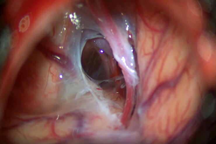
Minor’s Syndrome Surgical Intervention by Prof. Vincent Darrouzet
Minor’s disease, also called Superior Semicircular Canal Dehiscence (SSCD) or Minor’s syndrome, is a rare disorder of the inner ear that affects hearing and balance. The disease is characterized by…

How to Choose a Microscope for Reconstructive Surgery
Plastic and reconstructive surgery requires excellent visualization to repair intricate and fine structures. Oncological reconstructive surgery procedures are among the most delicate, including breast…

How to Remove Out-Of-Focus Blur and Improve Segmentation Accuracy
The specificity of fluorescence microscopy allows researchers to accurately observe and analyze biological processes and structures quickly and easily, even when using thick or large samples. However,…

Live-Cell Imaging Techniques
The understanding of complex and/or fast cellular dynamics is an important step for exploring biological processes. Therefore, today’s life science research is increasingly focused on dynamic…
The AI-Powered Pixel Classifier
Achieving reproducible results manually requires expertise and is tedious work. But now there is a way to overcome these challenges by speeding up this analysis to extract the real value of the image…

Applying AI and Machine Learning in Microscopy and Image Analysis
Prof. Emma Lundberg is a professor in cell biology proteomics at KTH Royal Institute of Technology, Sweden. She is also the director of the Cell Atlas, an integral part of the Swedish-based Human…

A New Method for Convenient and Efficient Multicolor Imaging
The technique combining hyperspectral unmixing and phasor analysis was developed to simplify the process of getting images from a sample labeled with multiple fluorophores. This aggregate method…

How to Select a Microscope for Cataract Surgery
What to consider in the selection of an ophthalmic microscope for cataract procedures. Bearing these aspects in mind will equip surgeons well for talks with manufacturer representatives. Many…
Optimal Visualization in Brain Surgery
This case study “Treatment of the Galassi type III arachnoid cyst with the M530 OHX surgical microscope from Leica Microsystems” documents the procedure step by step and shows the visualization…

Benefits of Combining STED and Lifetime
In this interview, Professor Alberto Diaspro talks about the advantages of the White Light Laser and the TauSTED capabilities of STELLARIS 8 STED. He speaks about his experience with the confocal…

How to use a Surgical Microscope as an Operating Room Nurse
Surgical microscopes play an essential role in the modern microsurgery procedures. It provides the surgeon, assistant and operating room staff with a magnified and illuminated high-quality image of…

Clinical Symposium on OCT-guided Cornea Surgery
In this recording Prof. Mehta from Singapore National Eye Centre and Prof. Fontana from Santa Maria
Nuova Hospital in Regio Emilia, Italy, share their expertise on corneal surgery. They present PK,…

Fluorescence Lifetime-based Imaging Gallery
Confocal microscopy relies on the effective excitation of fluorescence probes and the efficient collection of photons emitted from the fluorescence process. One aspect of fluorescence is the emission…

Towards Advanced Use of Intraoperative OCT in Cataract Surgery
In this White Paper, Dr. Rachid Tahiri shares his personal experience with the Leica EnFocus intraoperative OCT, the valuable features supporting smooth surgery and how it allows him to minimize…

How Clinical and Surgical Microscopes Support Otolaryngology
The number of ENT surgery procedures is increasing year after year. It is estimated that there will be over 21 million ENT procedures performed annually worldwide by 2022.
Dr. Duane Mol is the…

Physiology Image Gallery
Physiology is about the processes and functions within a living organism. Research in physiology focuses on the activities and functions of an organism’s organs, tissues, or cells, including the…

Live Cell Imaging Gallery
Live cell microscopy techniques are fundamental to get a better understanding of cellular and molecular function. Today, widefield microscopy is the most common technique used to visualize cell…
 and astrocytes (green) in a cortical spheroid derived from human induced pluripotent stem cells.")
Neuroscience Images
Neuroscience commonly uses microscopy to study the nervous system’s function and understand neurodegenerative diseases.
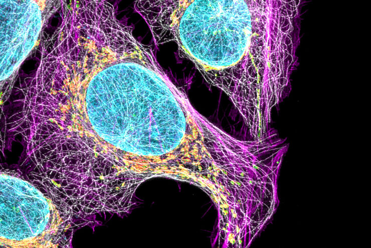
Cell Biology Image Gallery
Cell biology studies the structure, function and behavior of cells, including cell metabolism, cell cycle, and cell signaling. Fluorescence microscopes are an integral part of a cell biologist…

A Guide to Darkfield Microscopes
A darkfield microscope offers a way to view the structures of many types of biological specimens in greater contrast without the need of stains.

How to Quantify Changes in the Metabolic Status of Single Cells
Metabolic imaging based on fluorescence lifetime provides insights into the metabolic dynamics of cells, but its use has been limited as expertise in advanced microscopy techniques was needed.
Now,…

Putting Dynamic Live Cell Data into the Ultrastructural Context
With workflow Coral Life, searching for a needle in the haystack is a thing of the past. Take advantage of correlative light and electron microscopy to identify directly the right cell at the right…

AI in Microscopy Webinar
We demonstrate residual channel attention networks for restoring and enhancing volumetric time-lapse (4D) fluorescence microscopy data.

A Guide to Phase Contrast
A phase contrast light microscope offers a way to view the structures of many types of biological specimens in greater contrast without the need of stains.

Benefits of TauContrast to Image Complex Samples
In this interview, Dr. Timo Zimmermann talks about his experience with the application of TauSense tools and their potential for the investigation of demanding samples such as thick samples or…

Fast, High-quality Vitrification with the EM ICE High Pressure Freezer
The EM ICE High Pressure Freezer was developed with a unique freezing principle and uses only a single pressurization and cooling liquid: liquified nitrogen (LN2). This design enables three major…
How Augmented Reality is Transforming Vascular Neurosurgery
Augmented Reality is changing surgery, with new information helping to improve the precision and safety of procedures. This is especially true in vascular neurosurgery where Augmented Reality is…
Overcoming Ophthalmologic Surgery Challenges
Ophthalmology surgical procedures involving both the anterior and posterior segment can be particularly challenging. Good visualization is required to operate with precision and confidence.
Prof.…
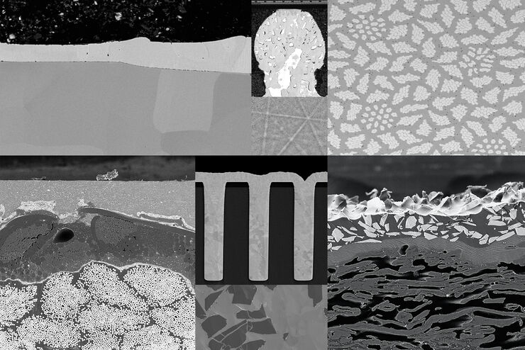
Ion Beam Milling Guide: Enhancing Surface Quality for High-Resolution Imaging and Analysis
In this article you can learn how to optimize the preparation quality of your samples by using the ion beam etching method with the EM TIC 3X ion beam milling machine. A short introduction of the…
