Corporate Communications

Leica Microsystems develops and manufactures microscopes and scientific instruments for the analysis of microstructures and nanostructures.
We offer scientific research and teaching material on the subjects of microscopy. The content is designed to support beginners, experienced practitioners and scientists alike in their everyday work and experiments. Explore interactive tutorials and application notes, discover the basics of microscopy as well as high-end technologies.
Follow us
 embedded in bulk vitreous ice (blue).")
Cryo-ET Sample Preparation: From Waffle Method to Serial Lift-Out
Cryo-ET sample preparation becomes more demanding when specimens are thicker, larger, or more complex. This webinar brings together four perspectives on how high-pressure freezing can be connected…
 and phalloidin (magenta), imaged using Viventis SCAPE; scale bar 50μm. Courtesy of Marina Cuenca and Heleen Jungen (Dayton lab), EMBL Barcelona.")
What’s the Best Organoid Imaging Approach for Early Drug Discovery?
Organoids and other complex in vitro models (CIVMs) are becoming increasingly important in early drug discovery and translational research, driven by the need for more predictive, human-relevant data…
 2D slices of a 1 mm diameter midbrain neural organoid stained with DAPI (blue, nuclear stain), β-tubulin (green, neuronal stain), and GFAP (red, astrocyte stain).")
Fast, High-Contrast Widefield Imaging of Optically Challenging Samples
Live‑cell imaging of large, complex biological samples often requires large fields of view, sub-cellular resolution, high-sensitivity, and fast acquisition – all while maintaining low illumination…

Dental Loupes vs Microscopes: Exploring Visualization Options in Dentistry
Dental professionals often ask: “Should I use dental loupes or invest in a microscope?” This article explores the key differences between dental microscopes and dental loupes, focusing on…

Spatial Proteomics Workflow in Blood Cancer (MPNs)
Megakaryocytes play a central role in the biology of myeloproliferative neoplasms (MPNs), yet their in vivo proteomic characterization remains a major challenge due to low abundance and disrupted…

Six Features to Consider when Choosing a Dental Microscope
The dental surgical microscope has become increasingly important for high-quality and successful dental medicine, particularly in the field of endodontics. A dentist can conduct micro-invasive…

Multiscale Imaging of Organoids: High Content to Light Sheet
Learn multiscale organoid imaging: fixed high content phenotyping, gentle dual view light sheet, and reproducible pipelines that turn 3D data into insights.

4 Key Benefits of 3D Digital Microscopy in Ophthalmic Surgery
3D digital visualization is rapidly transforming ophthalmic surgery. Modern 3D surgical microscopes enable surgeons to perform procedures using high-resolution digital displays rather than traditional…
, insulin SGs (orange), microtubules (red), nucleus (yellow), and plasma membrane (transparent).")
High-Pressure Freezing Protocols for Ultrastructural 3D EM
High pressure freezing (HPF) can help preserve hydrated cells and tissues close to their biological state at the moment of immobilization, supporting more reliable ultrastructural interpretation than…
Ultramicrotome UC Enuity in Practice: Stable 15 nm Sections at ZFE
After using the UCT and UC6 ultramicrotomes, Claudia Mayrhofer calls UC Enuity a leap in stability—so robust that vibrations and temperature shifts don’t spoil sections, even with multiple users. Auto…

Expert Techniques for Superior Visualization in Cataract Surgery
Join renowned ophthalmic surgeons, Dr. Hussein Almuhtaseb and Mr. Simon Madge, as they share their clinical expertise and real-world surgical strategies during the 2025 Online Cataract Surgery…

History, Developments and Trends of Microscopy in Cancer Research
Cancer is a global disease, with 18 million new cases diagnosed and 10 million cancer-related deaths worldwide in 2020. This burden is set to increase, with a projected increase in cases of ~55% by…

Researchers Insights: Microscopy in Cancer Research
Discover how imaging techniques are driving cancer research forward. In this issue, we present comprehensive multimodal studies using microscopy, as well as new directions in intraoperative cancer…

Predictive Service Prevents Downtime in Ghent
At the VIB BioImaging Core in Ghent, Belgium, researchers depend on Leica’s Stellaris 8 confocal microscope to explore the frontiers of biomedical science. When Leica’s RemoteCare system detected a…
, Tropomyosin (cardiomyocytes, red) and GFP (primordial cardiac layer, green).")
A Guide to Fluorescence Microscopy
Fluorescence microscopy uses the ability of fluorophores, dyes, or fluorescent proteins to emit light of a specific wavelength after being excited with light of a shorter wavelength. Biomolecules can…

Advances in Oncological Reconstructive Surgery
Decision making and patient care in oncological reconstructive surgery have considerably evolved in recent years. New surgical assistance technologies are helping surgeons push the boundaries of what…

Ultramicrotomy eBook: Targeting, Trimming & Alignment
Ultramicrotomy is evolving rapidly, and today’s microscopes demand high‑quality sections, precise targeting, and reproducible workflows. This eBook brings together expert application notes, automated…
 case.")
Flexibility and Efficiency in Minimally Invasive Spine Surgery
According to Prof. Alex Alfieri, Chief Physician and Head of clinic for Neurosurgery and Spinal surgery at the Cantonal Hospital Winterthur, Minimally invasive spine surgery (MISS) is transforming…
 labeled with membrane-permeable calcein, high-pressure frozen in salt water using EM ICE.")
High-Pressure Freezing for Organoids: Cryo CLEM & FIB Lift Out
Master cryo EM workflow steps for challenging 3D samples: when to choose HPF vs. plunge freezing, reproducible blotting/ice control, contamination aware transfers, Cryo CLEM 3D targeting in organoids,…
 and astrocytes (green) in a cortical spheroid derived from human induced pluripotent stem cells.")
Guide to Live-Cell Imaging
For a wide range of applications in various research fields of life science, live-cell imaging is an indispensable tool for visualizing cells in a state as close to in vivo, i.e. living and active, as…

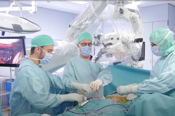
A Larger 3D Area in Focus for Neurosurgical and Ophthalmic Microscopes
Neurosurgeons and ophthalmologists deal with delicate structures, deep or narrow cavities and tiny structures with vitally important functions. Seeing a clear, large 3D area of the surgical field in…

Factors to Consider When Selecting a Research Microscope
An optical microscope is often one of the central devices in a life-science research lab. It can be used for various applications which shed light on many scientific questions. Thereby the…

Advanced Visualization in Head and Neck Reconstructive Surgery
PRS surgeons and maxillofacial specialists face unique challenges in reconstructive procedures, particularly when operating in deep, narrow cavities and working with delicate tissues. Achieving…
Advanced Visualization: Transforming Minimally Invasive Spine Surgery
Over the past decade, spine surgery has evolved rapidly with the adoption of minimally invasive surgery (MISS) techniques. These approaches reduce surgical trauma, speed up recovery, and help improve…

Advanced Visualization in ENT Surgery: Prof. Darrouzet’s Expert Insights
Otologic surgery demands exceptional precision when working within the confined spaces of the middle and inner ear. Procedures such as tympanoplasty, mastoidectomy, and cochlear implant placement…

AI meets Deep Visual Proteomics (DVP) to Advance Disease Research
In this webinar, Dr. Andreas Mund will introduce a cutting-edge platform that merges Deep Visual Proteomics (DVP) with AI-powered pathology models, enabling high-resolution mapping of key regions in…
 and co-stained for nuclear DNA (Hoechst 33342), microtubules (Alexa 555) and F-actin (ATTO 643). Image was captured on Mateo FL.")
Microscopy and AI Solutions for 2D Cell Culture
This eBook explores the integration of microscopy and AI technologies in 2D cell culture workflows. It highlights how traditional imaging methods—such as brightfield, phase contrast, and…
.")
Focus on Long-Term Imaging in 3D with Light Sheet Microscopy
Long-term 3D imaging reveals how complex multicellular systems grow and develop and how cells move and interact over time, unlocking critical insights into development, disease, and regeneration.…

Polarizing Microscope Image Gallery
How polarization microscope images can be used for analysis is shown in this gallery. Polarized light microscopy (also known as polarizing microscopy) is an important method for different fields and…

A Guide to C. elegans Research – Working with Nematodes
Efficient microscopy techniques for C. elegans research are outlined in this guide. As a widely used model organism with about 70% gene homology to humans, the nematode Caenorhabditis elegans (also…
