
Science Lab
Science Lab
Das Wissensportal von Leica Microsystems bietet Ihnen Wissens- und Lehrmaterial zu den Themen der Mikroskopie. Die Inhalte sind so konzipiert, dass sie Einsteiger, erfahrene Praktiker und Wissenschaftler gleichermaßen bei ihrem alltäglichen Vorgehen und Experimenten unterstützen. Entdecken Sie interaktive Tutorials und Anwendungsberichte, erfahren Sie mehr über die Grundlagen der Mikroskopie und High-End-Technologien - werden Sie Teil der Science Lab Community und teilen Sie Ihr Wissen!
Loading...

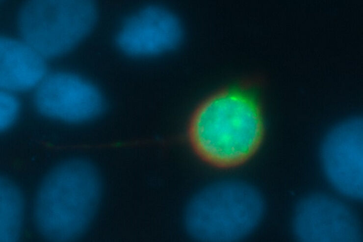
Die Rolle des Eisenstoffwechsels bei der Krebsentwicklung
Der Eisenstoffwechsel spielt eine Rolle bei der Entstehung und dem Fortschreiten von Krebs und beeinflusst die Immunreaktion. Zu verstehen, wie Eisen Krebs und das Immunsystem beeinflusst, kann die…
Loading...

Differential Interference Contrast (DIC) Microscopy
This article demonstrates how differential interference contrast (DIC) can be actually better than brightfield illumination when using microscopy to image unstained biological specimens.
Loading...
Going Beyond Deconvolution
Widefield fluorescence microscopy is often used to visualize structures in life science specimens and obtain useful information. With the use of fluorescent proteins or dyes, discrete specimen…
Loading...
 cells taken with phase contrast.")
Phase Contrast and Microscopy
This article explains phase contrast, an optical microscopy technique, which reveals fine details of unstained, transparent specimens that are difficult to see with common brightfield illumination.
Loading...

How to do a Proper Cell Culture Quick Check
In order to successfully work with mammalian cell lines, they must be grown under controlled conditions and require their own specific growth medium. In addition, to guarantee consistency their growth…
Loading...
FluoSync - a Fast & Gentle Method for Unmixing Multicolour Images
In this white paper, we focus on a fast and reliable method for obtaining high-quality multiplex images in fluorescence microscopy. FluoSync combines an existing method for hybrid unmixing with…
Loading...
, SPY-Actin (cyan), and SiR-Tubulin (magenta). Instant Computational Clearing (ICC) was applied.")
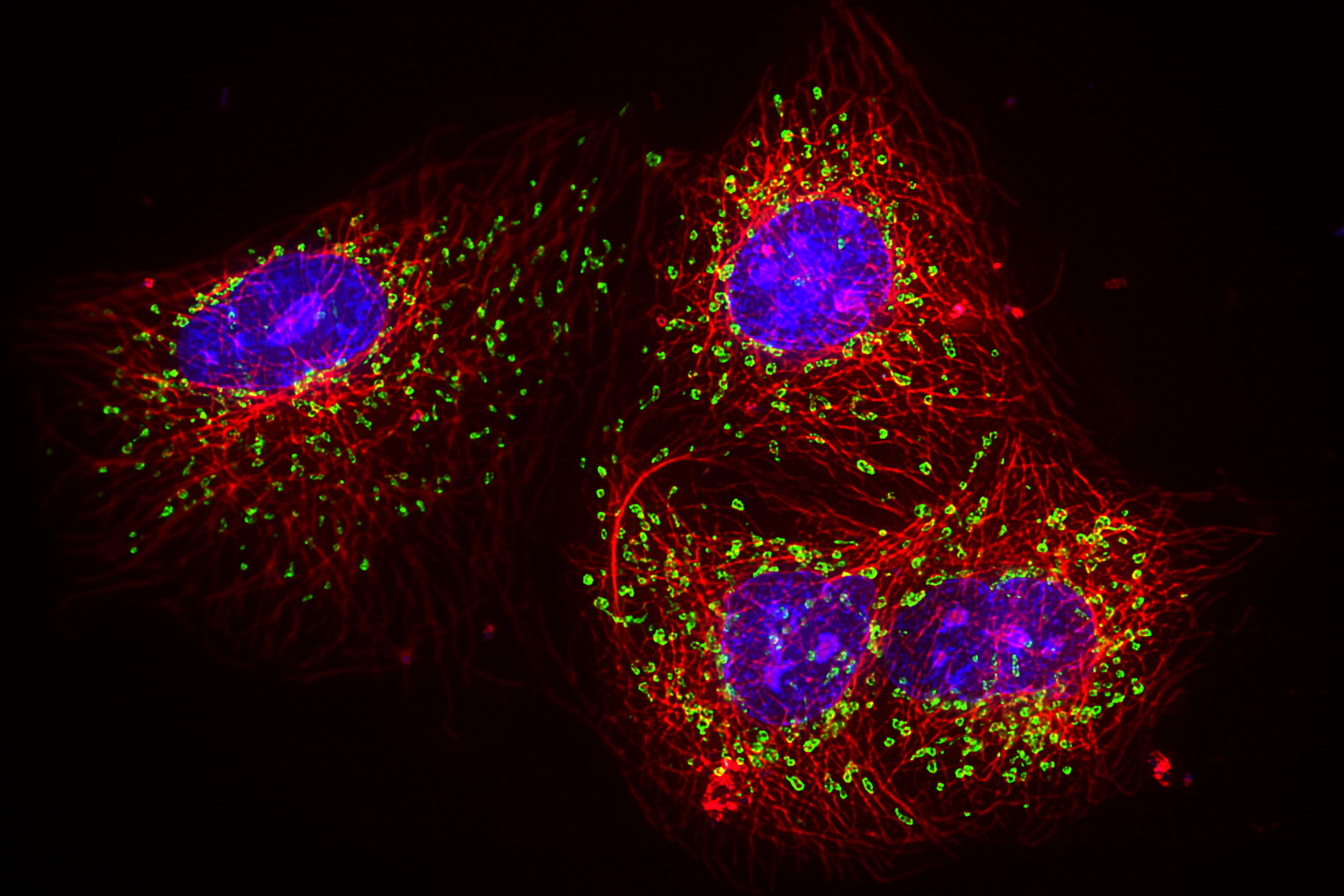
How to Perform Dynamic Multicolor Time-Lapse Imaging
Live-cell imaging sheds light on diverse cellular events. As many of these events have fast dynamics, the microscope imaging system must be fast enough to record every detail. One major advantage of…
Loading...
, the cis-golgi matrix protein GM130 (AF488, green), and the trans-golgi network membrane protein TGN46 (AF647, red).")
Golgi Organizational Changes in Response to Cell Stress
VIDEO ON DEMAND - In this episode of MicaCam, our special guest George Galea from EMBL Heidelberg will look at HeLa Kyoto cells treated with various chemotherapeutic agents to investigate their effect…
Loading...

Multiplexed Imaging Types, Benefits and Applications
Multiplexed imaging is an emerging and exciting way to extract information from human tissue samples by visualizing many more biomarkers than traditional microscopy. By observing many biomarkers…