
Life Science Research
Life Science Research
This is the place to expand your knowledge, research capabilities, and practical applications of microscopy in various scientific fields. Learn how to achieve precise visualization, image interpretation, and research advancements. Find insightful information on advanced microscopy, imaging techniques, sample preparation, and image analysis. Topics covered include cell biology, neuroscience, and cancer research with a focus on cutting-edge applications and innovations.

Inverted Microscopes for Grain Size Analysis: Three Factors to Consider
Microscopic steel grain size analysis is useful in determining the quality of steel alloys for a given purpose such as building bridges vs railroad rails. This webinar will describe the preparation of…

Analyzing Non-metallic Inclusions in Steel
Oftentimes we find ourselves caught up in tedious analyses by reticle and comparison chart, time-consuming double-evaluation according to several standards or subjective inspection results with a bias…

Computational Clearing - Enhance 3D Specimen Imaging
This webinar is designed to clarify crucial specifications that contribute to THUNDER Imagers' transformative visualization of 3D samples and improvements within a researcher's imaging-related…
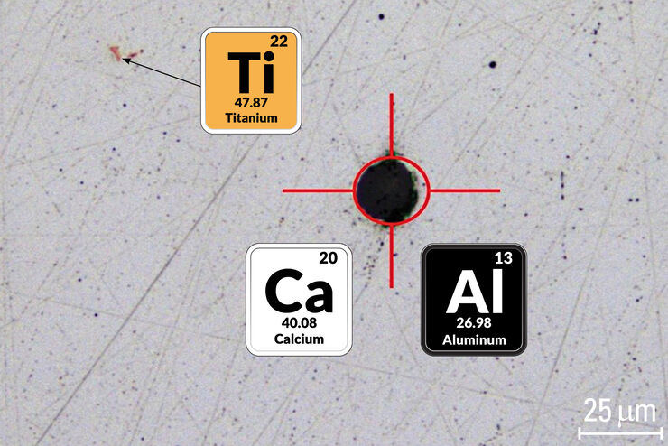
Visual and Chemical Analysis of Steel Microstructure: Faster Rating of Steel Quality
Simultaneous visual and chemical analysis of steel non-metallic inclusions with a 2-methods-in-1 solution, using optical microscopy and laser induced breakdown spectroscopy (LIBS), is described in…

Rate the Quality of Your Steel: Free Webinar and Report
This webinar and report describe optimal microscopy solutions for rating steel quality in terms of non-metallic inclusions and reviews the various international and regional standards concerning…

Guide to Microscopy in Cancer Research
Cancer is a complex and heterogeneous disease caused by cells deficient in growth regulation. Genetic and epigenetic changes in one or a group of cells disrupt normal function and result in…

THUNDER Imagers: High Performance, Versatility and Ease-of-Use for your Everyday Imaging Workflows
This webinar will showcase the versatility and performance of THUNDER Imagers in many different life science applications: from counting nuclei in retina sections and RNA molecules in cancer tissue…

Metallography – an Introduction
This article gives an overview of metallography and metallic alloy characterization. Different microscopy techniques are used to study the alloy microstructure, i.e., microscale structure of grains,…
Improve Cryo Electron Tomography Workflow
Leica Microsystems and Thermo Fisher Scientific have collaborated to create a fully integrated cryo-tomography workflow that responds to these research needs: Reveal cellular mechanisms at…
