Webinars
Take a look at our upcoming and on-demand webinars. Join us at one of our next events!
Take a look at all our upcoming congresses, exhibitions, webinars, and workshops and join us at one of our next events!
03
Jul
2025
显微切割技术在空间组学和药物开发中的应用探索
China
•
Webinar
03
Jul
2025
Filter articles
Tags
Products
Loading...

Cutting-Edge Imaging Techniques for GPCR Signaling
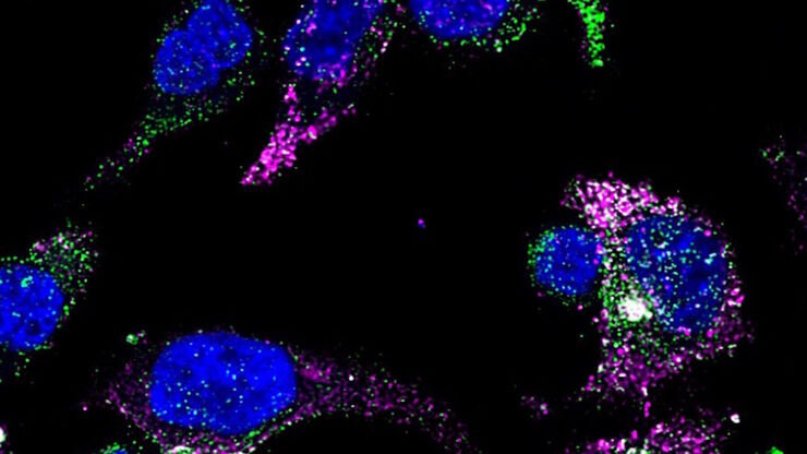
With this webinar on-demand enhance your pharmacological research with our webinar on GPCR signaling and explore cutting-edge imaging techniques that aim to understand how GPCR signaling translates…
Loading...

Revealing Neuronal Migration’s Molecular Secrets
Different approaches can be used to investigate neuronal migration to their niche in the developing brain. In this webinar, experts from The University of Oxford present the microscopy tools and…
Loading...

Accelerating Discovery for Multiplexed Imaging of Diverse Tissues
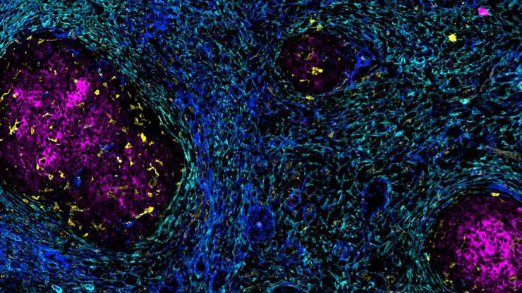
Explore IBEX: Open-source multiplexed imaging. Join the collaborative IBEX Imaging Community for optimized tissue processing, antibody selection, and human atlas construction.
Loading...

Notable AI-based Solutions for Phenotypic Drug Screening
Learn about notable optical microscope solutions for phenotypic drug screening using 3D-cell culture, both planning and execution, from this free, on-demand webinar.
Loading...

Understanding Tumor Heterogeneity with Protein Marker Imaging
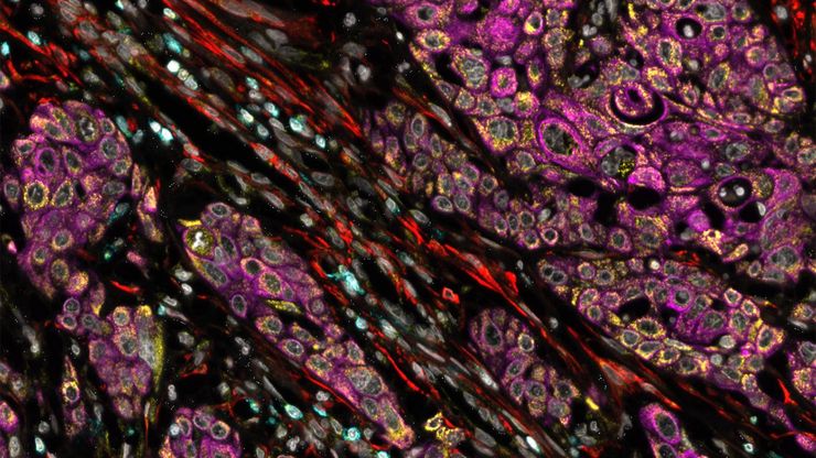
Explore tumor heterogeneity and immune cell dynamics. See how quantitative imaging analysis reveals spatial relationships and molecular insights crucial for advancing cancer research and therapeutics.
Loading...

Cell DIVE: Unraveling Pathways in Pancreatic Cancer
Unlock new insights into tumour-immune interactions with spatial biology. Join our webinar to learn how to label >60 biomarkers per tissue section and use advanced clustering and analysis techniques…
