
Science Lab
Science Lab
Bienvenue sur le portail de connaissances de Leica Microsystems. Vous y trouverez des recherches scientifiques et du matériel didactique sur le thème de la microscopie. Le portail aide les débutants, les praticiens expérimentés et les scientifiques dans leur travail quotidien et leurs expériences. Explorez les didacticiels interactifs et les notes d'application, découvrez les bases de la microscopie ainsi que les technologies de pointe. Faites partie de la communauté Science Lab et partagez votre expertise.
Filter articles
Tags
Type de publication
Produits
Loading...

Kinetochore Assembly during Mitosis with TauSTED on 3D
Three-dimensional organization of the mitotic spindle together with the distribution of CENP-C and BUB1 based on TauSTED with multiple STED lines (592, 660 and 775 nm) can provide insights…
Loading...

How to Quantify Changes in the Metabolic Status of Single Cells
Metabolic imaging based on fluorescence lifetime provides insights into the metabolic dynamics of cells, but its use has been limited as expertise in advanced microscopy techniques was needed.
Now,…
Loading...

Putting Dynamic Live Cell Data into the Ultrastructural Context
With workflow Coral Life, searching for a needle in the haystack is a thing of the past. Take advantage of correlative light and electron microscopy to identify directly the right cell at the right…
Loading...

AI in Microscopy Webinar
We demonstrate residual channel attention networks for restoring and enhancing volumetric time-lapse (4D) fluorescence microscopy data.
Loading...

20 Years of Leica Laser Microdissection
Phenotype-genotype correlations are key for insight. From Eye to Insight is therefore fitting perfectly to Leica Microsystems and in particular to laser microdissection. Laser Microdissection, also…
Loading...

A Guide to Phase Contrast
A phase contrast light microscope offers a way to view the structures of many types of biological specimens in greater contrast without the need of stains.
Loading...

Benefits of TauContrast to Image Complex Samples
In this interview, Dr. Timo Zimmermann talks about his experience with the application of TauSense tools and their potential for the investigation of demanding samples such as thick samples or…
Loading...
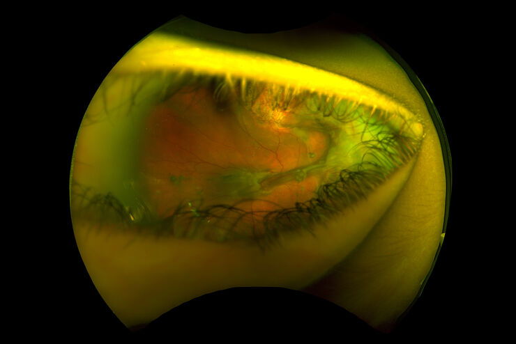
Intraoperative OCT-Assisted Surgical Management of Proliferative Vitreoretinopathy
Proliferative vitreoretinopathy (PVR) is a plague to patients and their surgeons after recent rhegmatogenous retinal detachment (RD). Despite excellent initial surgical outcomes, it is the most common…
Loading...

Fast, High-quality Vitrification with the EM ICE High Pressure Freezer
The EM ICE High Pressure Freezer was developed with a unique freezing principle and uses only a single pressurization and cooling liquid: liquified nitrogen (LN2). This design enables three major…
