
Industrial Microscopy
Industrial Microscopy
Dive deep into detailed articles and webinars focusing on efficient inspection, optimized workflows, and ergonomic comfort in industrial and pathological contexts. Topics covered include quality control, materials analysis, microscopy in pathology, among many others. This is the place where you get valuable insights into using cutting-edge technologies for improving precision and efficiency in manufacturing processes as well as accurate pathological diagnosis and research.

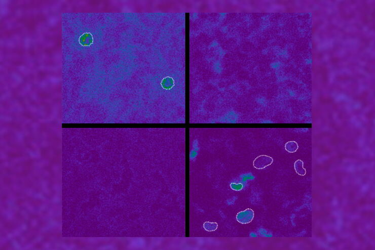
Simplifying Complex Fluorescence Multiwell Plate Assays
Apoptosis, or programmed cell death, occurs during organism embryo development to eliminate unwanted cells and during healing in adults to rid the body of damaged cells and help prevent cancer.…

Efficient Long-term Time-lapse Microscopy
When doing time-lapse microscopy experiments with spheroids, there are certain challenges which can arise. As the experiments can last for several days, prolonged sample survival must be achieved…

How to Choose a Microscope for Reconstructive Surgery
Plastic and reconstructive surgery requires excellent visualization to repair intricate and fine structures. Oncological reconstructive surgery procedures are among the most delicate, including breast…

How to Prepare your Specimen for Immunofluorescence Microscopy
Immunofluorescence (IF) specimen preparations can be analyzed by various fluorescence microscopy techniques, depending on the research application. IF is indispensable for researchers using a simple…

How Industrial Applications Benefit from Fluorescence Microscopy
See what you can do with fluorescence microscopy for industrial and materials science applications from this webinar. Fluorescence is well known for biological applications, however, almost all…
Spectroscopic Evaluation of Red Blood Cells
Hemoglobinopathies are a major healthcare problem. This study presents a possible diagnostic tool for thalassemia which is based on confocal spectroscopy. This approach exploits spectral detection and…
How Augmented Reality is Transforming Vascular Neurosurgery
Augmented Reality is changing surgery, with new information helping to improve the precision and safety of procedures. This is especially true in vascular neurosurgery where Augmented Reality is…

How FLIM Microscopy Helps to Detect Microplastic Pollution
The use of autofluorescence in biological samples is a widely used method to gain detailed knowledge about systems or organisms. This property is not only found in biological systems, but also…
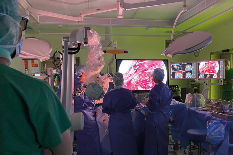
Neurosurgery with Heads-up Display
In the following video interviews Prof. Dr. Raphael Guzman, Vice Chairman of the Department of Neurosurgery at the University Hospital in Basel, Switzerland, talks about his experience in heads-up…
