

Science Lab
Science Lab
Willkommen auf dem Wissensportal von Leica Microsystems. Hier finden Sie wissenschaftliches Forschungs- und Lehrmaterial rund um das Thema Mikroskopie. Das Portal unterstützt Anfänger, erfahrene Praktiker und Wissenschaftler gleichermaßen bei ihrer täglichen Arbeit und ihren Experimenten. Erkunden Sie interaktive Tutorials und Anwendungshinweise, entdecken Sie die Grundlagen der Mikroskopie ebenso wie High-End-Technologien. Werden Sie Teil der Science Lab Community und teilen Sie Ihr Fachwissen.
Filter articles
Tags
Beitragstyp
Produkte
Loading...
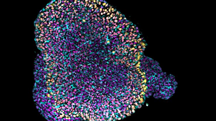
Volumetric Imaging of Cleared Organoids with Light Sheet
Cleared organoid imaging with Viventis Deep light sheet: rapid, high-resolution imaging of large organoids throughout their full volume.
Loading...
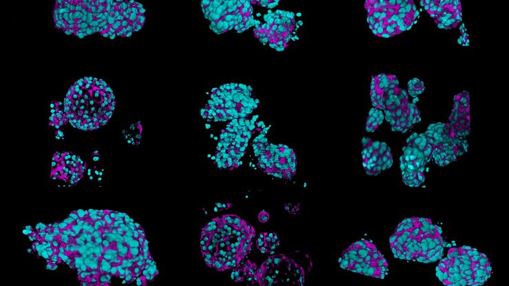
What’s the Best Organoid Imaging Approach for Early Drug Discovery?
Organoids and other complex in vitro models (CIVMs) are becoming increasingly important in early drug discovery and translational research, driven by the need for more predictive, human-relevant data…
Loading...
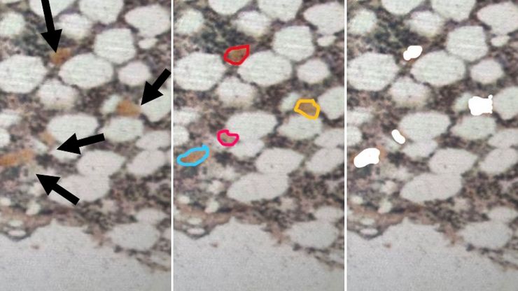
Spatial Proteomics Workflow in Blood Cancer (MPNs)
Megakaryocytes play a central role in the biology of myeloproliferative neoplasms (MPNs), yet their in vivo proteomic characterization remains a major challenge due to low abundance and disrupted…
Loading...

History, Developments and Trends of Microscopy in Cancer Research
Cancer is a global disease, with 18 million new cases diagnosed and 10 million cancer-related deaths worldwide in 2020. This burden is set to increase, with a projected increase in cases of ~55% by…
Loading...

How to Select the Right Measurement Microscope
With a measurement microscope, users can measure the size and dimensions of sample features in both 2D and 3D, something crucial for inspection, QC, failure analysis, and R&D. However, choosing the…
Loading...
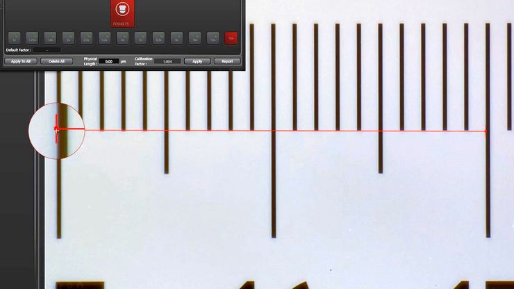
Microscope Calibration for Measurements: Why and How You Should Do It
Microscope calibration ensures accurate and consistent measurements for inspection, quality control (QC), failure analysis, and research and development (R&D). Calibration steps are described in this…
Loading...
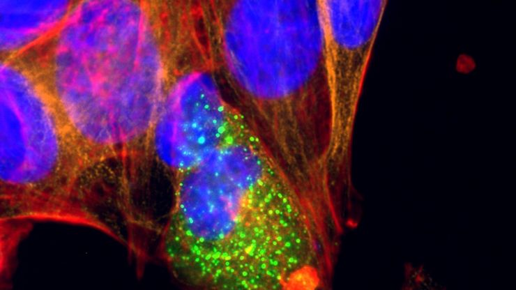
Microscopy and AI Solutions for 2D Cell Culture
This eBook explores the integration of microscopy and AI technologies in 2D cell culture workflows. It highlights how traditional imaging methods—such as brightfield, phase contrast, and…
Loading...
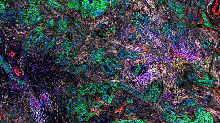
AI-Powered Hi-Plex Spatial Analysis Tools for Breast Cancer Research
Breast cancer (BC) is the leading cause of cancer-related deaths in women. Investigating the tumor microenvironment (TME) is crucial to elucidate the mechanisms of tumor progression. Systematic…
Loading...
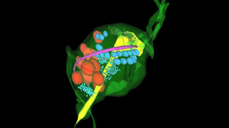
Volume EM and AI Image Analysis
The article outlines a detailed workflow for studying biological tissues in three dimensions using volume-scanning electron microscopy (volume-SEM) combined with AI-assisted image analysis. The focus…


