
Science Lab
Science Lab
Willkommen auf dem Wissensportal von Leica Microsystems. Hier finden Sie wissenschaftliches Forschungs- und Lehrmaterial rund um das Thema Mikroskopie. Das Portal unterstützt Anfänger, erfahrene Praktiker und Wissenschaftler gleichermaßen bei ihrer täglichen Arbeit und ihren Experimenten. Erkunden Sie interaktive Tutorials und Anwendungshinweise, entdecken Sie die Grundlagen der Mikroskopie ebenso wie High-End-Technologien. Werden Sie Teil der Science Lab Community und teilen Sie Ihr Fachwissen.
Filter articles
Tags
Beitragstyp
Produkte
Loading...
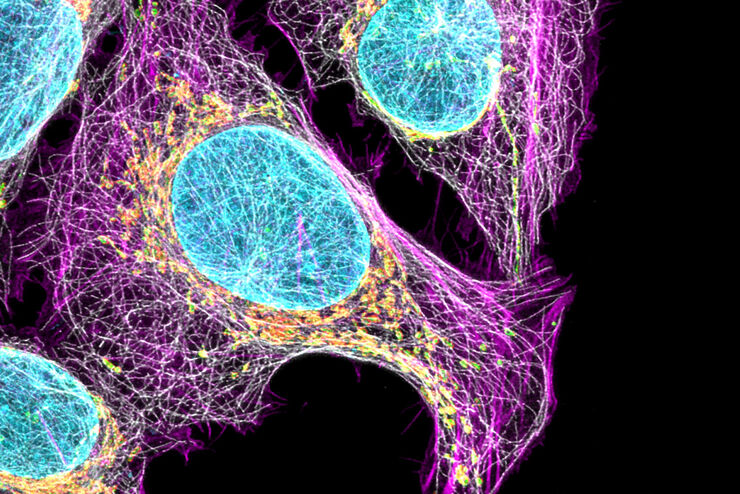
Cell Biology Image Gallery
Cell biology studies the structure, function and behavior of cells, including cell metabolism, cell cycle, and cell signaling. Fluorescence microscopes are an integral part of a cell biologist…
Loading...

Developmental Biology Image Gallery
Developmental biology explores the development of complex organisms from the embryo to adulthood to understand in detail the origins of disease. This category of the gallery shows images about…
Loading...

Image Gallery: THUNDER Imager
To help you answer important scientific questions, THUNDER Imagers eliminate the out-of-focus blur that clouds the view of thick samples when using camera-based fluorescence microscopes. They achieve…
Loading...
Augmented Reality (AR) Fluorescence Image Gallery
Building on a decade of leadership in fluorescence imaging technology, GLOW800 AR fluorescence is the first of many modalities based on the proprietary GLOW AR platform.
The sophisticated imaging…
Loading...
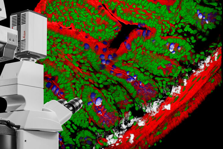
DIVE Multiphoton Microscope Image Gallery
Today’s life science research focusses on complex biological processes, such as the causes of cancer and other human diseases. A deep look into tissues and living specimens is vital to understanding…
Loading...
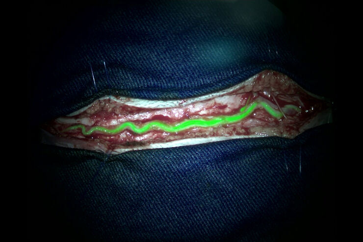
Digital Microscopy in Forensics
Forensic experts work with a broad range of microscopes to examine evidence from firearms and tool marks, documents, forensic or legal medicine, hair and fibers as well as glass and paint. Digital…
Loading...
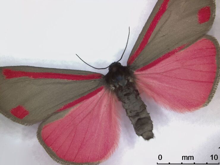
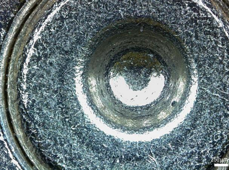
Life Science Imaging with DVM6 Digital Microscope
Digital microscopes can be a great help in life science applications such as the documentation in botany, entomology studies and crop science, or the digitization of museum collections. The image…
