
Science Lab
Science Lab
Willkommen auf dem Wissensportal von Leica Microsystems. Hier finden Sie wissenschaftliches Forschungs- und Lehrmaterial rund um das Thema Mikroskopie. Das Portal unterstützt Anfänger, erfahrene Praktiker und Wissenschaftler gleichermaßen bei ihrer täglichen Arbeit und ihren Experimenten. Erkunden Sie interaktive Tutorials und Anwendungshinweise, entdecken Sie die Grundlagen der Mikroskopie ebenso wie High-End-Technologien. Werden Sie Teil der Science Lab Community und teilen Sie Ihr Fachwissen.
Filter articles
Tags
Beitragstyp
Produkte
Loading...

How does an Automated Rating Solution for Steel Inclusions Work?
The rating of non-metallic inclusions (NMIs) to determine steel quality is critical for many industrial applications. For an efficient and cost-effective steel quality evaluation, an automated NMI…
Loading...

Perform Microscopy Analysis for Pathology Ergonomically and Efficiently
The main performance features of a microscope which are critical for rapid, ergonomic, and precise microscopic analysis of pathology specimens are described in this article. Microscopic analysis of…
Loading...

How to Conduct Standard-Compliant Analysis of Non-Metallic Inclusions in Steel
This webinar will provide an overview of the significance of non-metallic inclusions in steel and outline the important global standards for rating the quality of steel and difficulties that arise in…
Loading...

Plastische und rekonstruktive Chirurgie: Warum ein Mikroskop benutzen?
Verfahren der plastischen und rekonstruktiven Chirurgie können heikel sein. Visualisierungslösungen spielen eine wichtige Rolle, da sie es ermöglichen, die Operation unter bestmöglichen Bedingungen…
Loading...
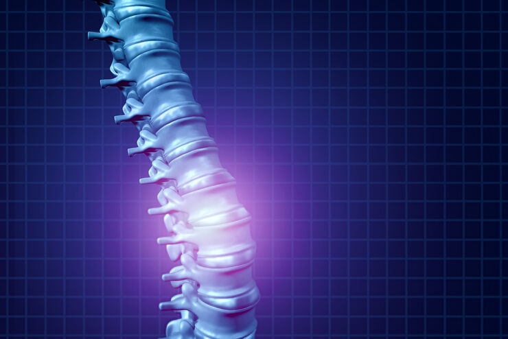
Minimally Invasive Spine Surgery: Improving Precision and Accuracy with Microscopes
Spine surgery is extremely delicate and requires extensive training and experience. Innovative visualization technologies can also help achieve better outcomes allowing to see more and have a clearer…
Loading...
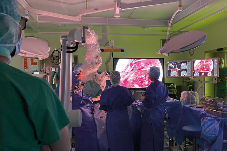
Neurosurgery with Heads-up Display
In the following video interviews Prof. Dr. Raphael Guzman, Vice Chairman of the Department of Neurosurgery at the University Hospital in Basel, Switzerland, talks about his experience in heads-up…
Loading...

Workflows and Instrumentation for Cryo-electron Microscopy
Cryo-electron microscopy is an increasingly popular modality to study the structures of macromolecular complexes and has enabled numerous new insights in cell biology. In recent years, cryo-electron…
Loading...

Modellorganismen in der Forschung
Modellorganismen sind Spezies, mit denen Forscher bestimmte biologische Vorgänge untersuchen. Sie haben genetische Ähnlichkeiten mit Menschen und werden häufig in Forschungsbereichen wie Genetik,…
Loading...

An Introduction to Computational Clearing
Many software packages include background subtraction algorithms to enhance the contrast of features in the image by reducing background noise. The most common methods used to remove background noise…
