

Science Lab
Science Lab
Willkommen auf dem Wissensportal von Leica Microsystems. Hier finden Sie wissenschaftliches Forschungs- und Lehrmaterial rund um das Thema Mikroskopie. Das Portal unterstützt Anfänger, erfahrene Praktiker und Wissenschaftler gleichermaßen bei ihrer täglichen Arbeit und ihren Experimenten. Erkunden Sie interaktive Tutorials und Anwendungshinweise, entdecken Sie die Grundlagen der Mikroskopie ebenso wie High-End-Technologien. Werden Sie Teil der Science Lab Community und teilen Sie Ihr Fachwissen.
Filter articles
Tags
Beitragstyp
Produkte
Loading...
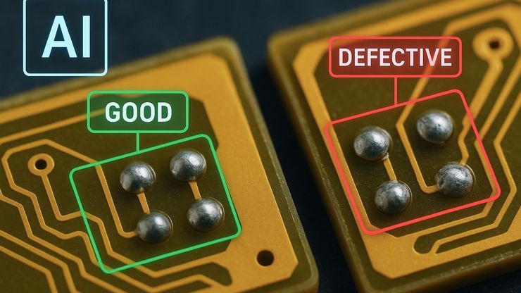
AI Image Analysis for Reliable Defect Detection
AI-based visual inspection with microscopy helps make quality control in industrial environments efficient by reducing manual inspection effort concerning defect detection, classification, and…
Loading...

An Overview of Artificial Intelligence (AI) and Machine Learning
AI is rapidly changing industrial inspection, quality control, and manufacturing workflows. But not all AI technologies work the same way. From machine learning and deep learning to the newer concept…
Loading...

Why the Future of Microscopic Inspection Is AI-Assisted, Not Fully Automated
The next wave of QC productivity gains will not come from building more automated inspection lines. Instead, it will come from augmenting millions of inspection decisions that are made manually every…
Loading...

How to Select the Right Measurement Microscope
With a measurement microscope, users can measure the size and dimensions of sample features in both 2D and 3D, something crucial for inspection, QC, failure analysis, and R&D. However, choosing the…
Loading...
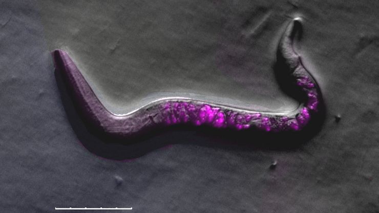
A Guide to C. elegans Research – Working with Nematodes
Efficient microscopy techniques for C. elegans research are outlined in this guide. As a widely used model organism with about 70% gene homology to humans, the nematode Caenorhabditis elegans (also…
Loading...
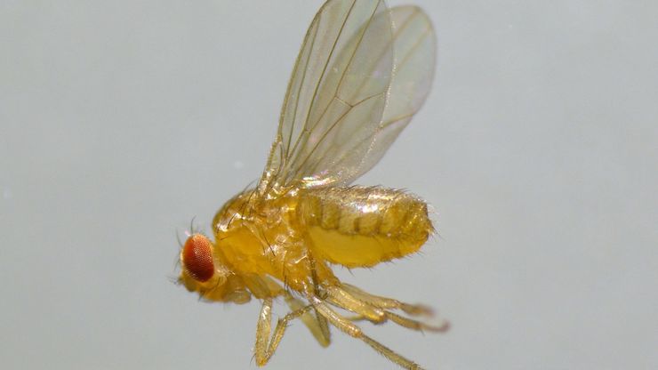
A Guide to Using Microscopy for Drosophila (Fruit Fly) Research
The fruit fly, typically Drosophila melanogaster, has been used as a model organism for over a century. One reason is that many disease-related genes are shared between Drosophila and humans. It is…
Loading...
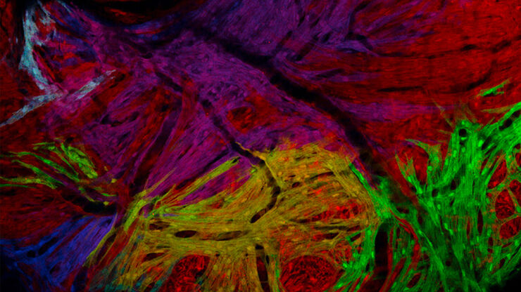
Zebrafisch-Forschung
Für optimale Ergebnisse während der Bewertung, Sortierung, Manipulation und Bildgebung von Modellorganismen ist es entscheidend feine Details und Strukturen genauestens zu erkennen. Das bildet die…
Loading...
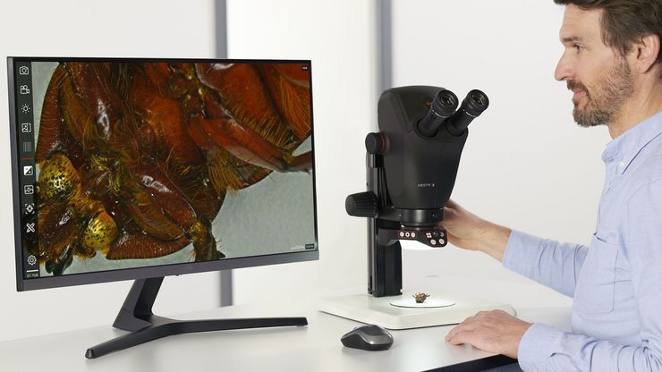
Präpariermikroskope
Wenn Sie Präparationen durchführen, verbringen Sie oft viele Stunden an den Okularen eines Präpariermikroskops. Bei Leica Microsystems können Sie aus einer Vielzahl von Mikroskopen und einem…
Loading...
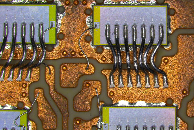
Top Challenges for Visual Inspection
This article discusses the challenges encountered when performing visual inspection and rework using a microscope. Using the right type of microscope and optical setup is paramount in order to…


