
Science Lab
Science Lab
Willkommen auf dem Wissensportal von Leica Microsystems. Hier finden Sie wissenschaftliches Forschungs- und Lehrmaterial rund um das Thema Mikroskopie. Das Portal unterstützt Anfänger, erfahrene Praktiker und Wissenschaftler gleichermaßen bei ihrer täglichen Arbeit und ihren Experimenten. Erkunden Sie interaktive Tutorials und Anwendungshinweise, entdecken Sie die Grundlagen der Mikroskopie ebenso wie High-End-Technologien. Werden Sie Teil der Science Lab Community und teilen Sie Ihr Fachwissen.
Filter articles
Tags
Beitragstyp
Produkte
Loading...

Image Gallery: THUNDER Imager
To help you answer important scientific questions, THUNDER Imagers eliminate the out-of-focus blur that clouds the view of thick samples when using camera-based fluorescence microscopes. They achieve…
Loading...

Information about Consumables for Laser Microdissection (LMD)
Types of consumables for Leica laser microdissection (LMD) systems, covering from basic to highly specialized applications, are described here. The most common ones are glass membrane slides in…
Loading...

An Introduction to Computational Clearing
Many software packages include background subtraction algorithms to enhance the contrast of features in the image by reducing background noise. The most common methods used to remove background noise…
Loading...

Microscopy in Virology
The coronavirus SARS-CoV-2, causing the Covid-19 disease effects our world in all aspects. Research to find immunization and treatment methods, in other words to fight this virus, gained highest…
Loading...

Computational Clearing - Enhance 3D Specimen Imaging
This webinar is designed to clarify crucial specifications that contribute to THUNDER Imagers' transformative visualization of 3D samples and improvements within a researcher's imaging-related…
Loading...

THUNDER Imagers: High Performance, Versatility and Ease-of-Use for your Everyday Imaging Workflows
This webinar will showcase the versatility and performance of THUNDER Imagers in many different life science applications: from counting nuclei in retina sections and RNA molecules in cancer tissue…
Loading...
 and YOYO 1 iodide (Nucleus).")
Real Time Images of 3D Specimens with Sharp Contrast Free of Haze
THUNDER Imagers deliver in real time images of 3D specimens with sharp contrast, free of the haze or out-of-focus blur typical of widefield systems. They can even image clearly places deep inside a…
Loading...
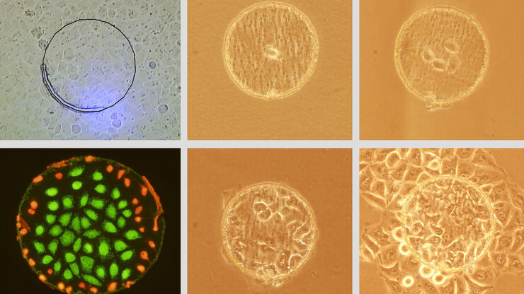
Live Cell Isolation by Laser Microdissection
Laser microdissection is a tool for the isolation of homogenous cell populations from their native niches in tissues to downstream molecular assays. Beside its routine use for fixed tissue sections,…
Loading...

Mission Impossible Accomplished: Tunable Colors for Non-descanning Detection
Leica Microsystems’ 4Tune detector, the key component of the SP8 DIVE Deep In Vivo Explorer, provides spectrally tunable image recording with non-descanning detection. An innovative solution for…
