Filter articles
タグ
製品
Loading...

Improving Zebrafish-Embryo Screening with Fast, High-Contrast Imaging
Discover from this article how screening of transgenic zebrafish embryos is boosted with high-speed, high-contrast imaging using the DM6 B microscope, ensuring accurate targeting for developmental…
Loading...

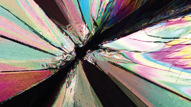
A Guide to Polarized Light Microscopy
Polarized light microscopy (POL) enhances contrast in birefringent materials and is used in geology, biology, and materials science to study minerals, crystals, fibers, and plant cell walls.
Loading...

顕微鏡を知る:被写界深度
顕微鏡において被写界深度は、凹凸の変化が⼤きい構造を持つ試料をピントがあったシャープに観察・撮像するために重要なパラメータです。被写界深度は、開⼝数、解像度、倍率の相関関係によって決定され、解像度とパラメータは反⽐例の関係にあります。被写界深度と解像度のバランスが最適になるように調整することができる顕微鏡もあります。
Loading...

Deep Visual Proteomics Provides Precise Spatial Proteomic Information
Despite the availability of imaging methods and mass spectroscopy for spatial proteomics, a key challenge that remains is correlating images with single-cell resolution to protein-abundance…
Loading...

What is Empty Magnification and How can Users Avoid it
The phenomenon of “empty magnification”, which can occur while using an optical, light, or digital microscope, and how it can be avoided is explained in this article. The performance of an optical…
Loading...

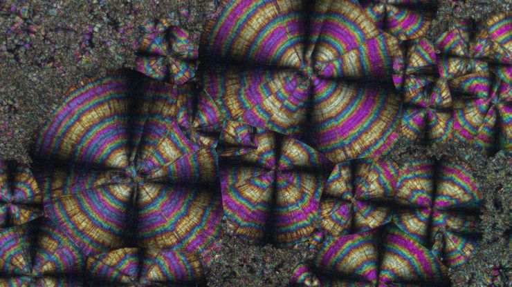
The Polarization Microscopy Principle
Polarization microscopy is routinely used in the material and earth sciences to identify materials and minerals on the basis of their characteristic refractive properties and colors. In biology,…
Loading...

神経細胞移動の分子的秘密を解き明かす
発達中の脳における特定部位への神経細胞の移動を調べるには、さまざまなアプローチを用いることができます。このウェビナーでは、オックスフォード大学の専門家が、神経発達過程における大脳皮質の機能層への神経細胞移動の分子メカニズムを解明するために使用している顕微鏡ツールとアッセイについて紹介します。これらのプロセスを理解することは、健全な脳の発達の理解を深め、神経発達障害の治療法を改善する可能性につながり…
Loading...

Empowering Spatial Biology with Open Multiplexing and Cell DIVE
Spatial biology and multiplexed imaging workflows have become important in immuno-oncology research. Many researchers struggle with study efficiency, even with effective tools and protocols. Here, we…
Loading...

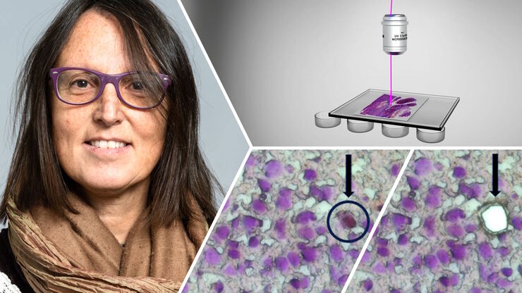
How did Laser Microdissection enable Pioneering Neuroscience Research?
Dr. Marta Paterlini, a Senior Scientist at the Karolinska Institute, shares her experience of using laser microdissection (LMD) in groundbreaking research into adult human neurogenesis and offers…

