
Life Science Research
Life Science Research
This is the place to expand your knowledge, research capabilities, and practical applications of microscopy in various scientific fields. Learn how to achieve precise visualization, image interpretation, and research advancements. Find insightful information on advanced microscopy, imaging techniques, sample preparation, and image analysis. Topics covered include cell biology, neuroscience, and cancer research with a focus on cutting-edge applications and innovations.

Sample Preparation for EM: A Practical Guide to Coating and Freeze-Fracturing
From coatings done in a low-vacuum sputter coating machine at room-temperature to those done in high-vacuum and even at cryogenic temperatures, Leica coating solutions cover a large range of needs.…

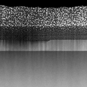
High Resolution Array Tomography with Automated Serial Sectioning
The optimization of high resolution, 3-dimensional (3D), sub-cellular structure analysis with array tomography using an automated serial sectioning solution, achieving a high section density on the…

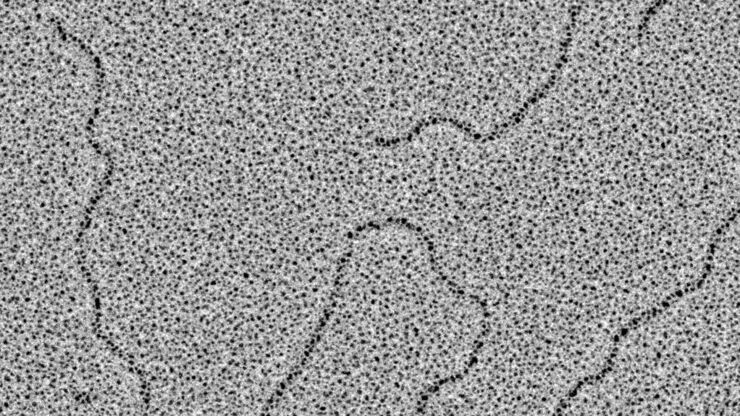
Visualization of DNA Molecules
Precise low angle rotary shadowing with heavy metals (like platinum) can be used in transmission electron microscopy (TEM) to observe molecular details of objects previously absorbed on a thin, low…
Each Atom Counts: Protect Your Samples Prior to FIB Processing
Application Note for Leica EM ACE600 - Focused ion beam (FIB) technology has become an indispensable tool for site-specific TEM sample preparation. It allows to extract electron transparent specimens…
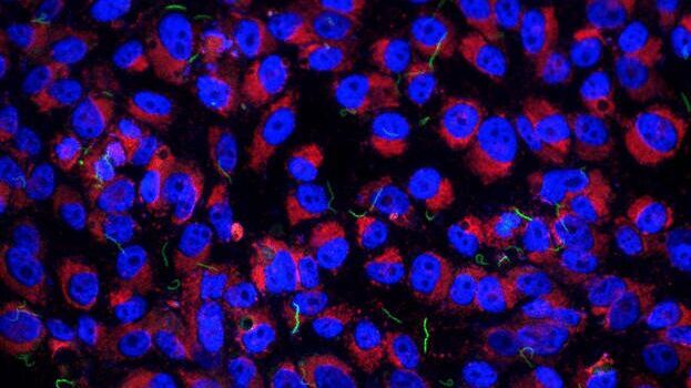
Imaging of Host Cell-bacteria Interactions using Correlative Microscopy under Cryo-conditions
Pathogenic bacteria have developed intriguing strategies to establish and promote infections in their respective hosts. Most bacterial pathogens initiate infectious diseases by adhering to host cells…
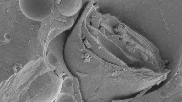
Brief Introduction to Freeze Fracture and Etching
Freeze fracture describes the technique of breaking a frozen specimen to reveal internal structures. Freeze etching is the sublimation of surface ice under vacuum to reveal details of the fractured…
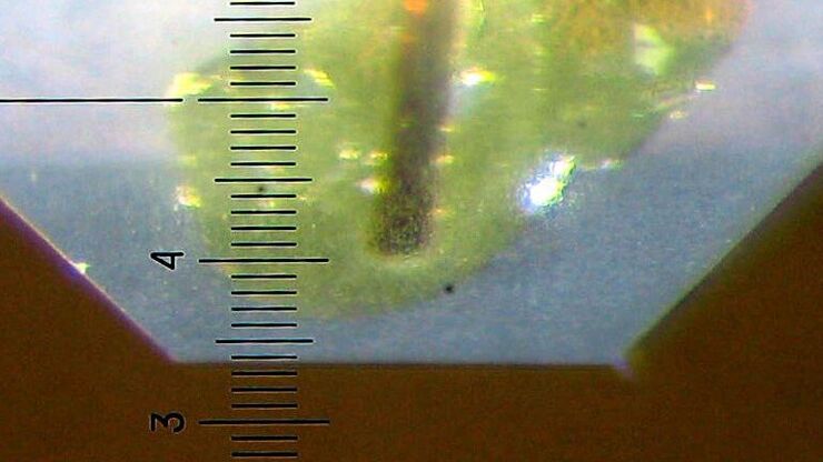
Brief Introduction to Specimen Trimming
Before ultrathin sectioning a sample with an ultramicrotome it has to be pre-prepared. For this pre-preparation, special attention must be paid to the sample size (size of the section), location of…
Glycerol Spraying/Platinum Low Angle Rotary Shadowing of DNA with the Leica EM ACE600 e-beam
Glycerol spraying/low angle rotary shadowing (Aebi and Baschong, 2006) is a preparation technique used in biology to visualize structures yielding insufficent contrast with other techniques, due to…

Brief Introduction to Coating Technology for Electron Microscopy
Coating of samples is required in the field of electron microscopy to enable or improve the imaging of samples. Creating a conductive layer of metal on the sample inhibits charging, reduces thermal…
