

Science Lab
Science Lab
Willkommen auf dem Wissensportal von Leica Microsystems. Hier finden Sie wissenschaftliches Forschungs- und Lehrmaterial rund um das Thema Mikroskopie. Das Portal unterstützt Anfänger, erfahrene Praktiker und Wissenschaftler gleichermaßen bei ihrer täglichen Arbeit und ihren Experimenten. Erkunden Sie interaktive Tutorials und Anwendungshinweise, entdecken Sie die Grundlagen der Mikroskopie ebenso wie High-End-Technologien. Werden Sie Teil der Science Lab Community und teilen Sie Ihr Fachwissen.
Filter articles
Tags
Beitragstyp
Produkte
Loading...

The Principles of White Light Laser Confocal Microscopy
The perfect light source for confocal microscopes in biomedical applications has sufficient intensity, tunable color and is pulsed for use in lifetime fluorescence. Furthermore, it should offer means…
Loading...

Introduction to Live-Cell Imaging
The understanding of complex and fast cellular dynamics is an important step to get insight into biological processes. Therefore, today’s life science research more and more demands studying…
Loading...
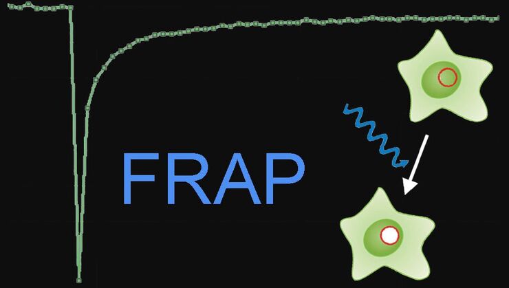
Fluorescence Recovery after Photobleaching (FRAP) and its Offspring
FRAP (Fluorescence recovery after photobleaching) can be used to study cellular protein dynamics: For visualization the protein of interest is fused to a fluorescent protein or a fluorescent dye. A…
Loading...
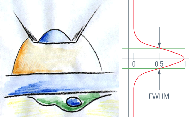
Confocal Optical Section Thickness
Confocal microscopes are employed to optically slice comparably thick samples.


