
Science Lab
Science Lab
Willkommen auf dem Wissensportal von Leica Microsystems. Hier finden Sie wissenschaftliches Forschungs- und Lehrmaterial rund um das Thema Mikroskopie. Das Portal unterstützt Anfänger, erfahrene Praktiker und Wissenschaftler gleichermaßen bei ihrer täglichen Arbeit und ihren Experimenten. Erkunden Sie interaktive Tutorials und Anwendungshinweise, entdecken Sie die Grundlagen der Mikroskopie ebenso wie High-End-Technologien. Werden Sie Teil der Science Lab Community und teilen Sie Ihr Fachwissen.
Filter articles
Tags
Beitragstyp
Produkte
Loading...
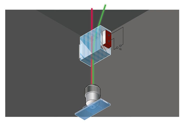
Primary Beam Splitting Devices for Confocal Microscopes
Current fluorescence microscopy employs incident illumination which requires separation of illumination and emission light. The classical device performing this separation is a color-dependent beam…
Loading...

Was macht das Pinhole im konfokalen Mikroskop?
Diese kurze einführende Schrift soll die Bedeutung des Pinholes erläutern und ist für Leser gedacht, die nicht allzu viel Zeit mit der Theorie und den Details der konfokalen Mikroskopie verbringen…
Loading...

Studying Caenorhabditis elegans (C. elegans)
Find out how you can image and study C. elegans roundworm model organisms efficiently with a microscope for developmental biology applications from this article.
Loading...

Vom Licht zur Erleuchtung: Sensoren und Messverfahren in der konfokalen Mikroskopie
In diesem Beitrag werden die wichtigsten Sensoren kurz vorgestellt, die in der konfokalen Mikroskopie verwendet werden. Mit konfokaler Mikroskopie ist hier „True Confo-cal Scanning“ gemeint, also das…
Loading...

Confocal and Light Sheet Imaging
Optical imaging instrumentation can magnify tiny objects, zoom in on distant stars and reveal details that are invisible to the naked eye. But it notoriously suffers from an annoying problem: the…
Loading...

Acousto Optics in True Confocal Spectral Microscope Systems
Acousto-optical elements have successfully replaced planar filters in many positions. The white confocal, regarded as the fully spectrally tunable confocal microscope, was not possible without this…
Loading...
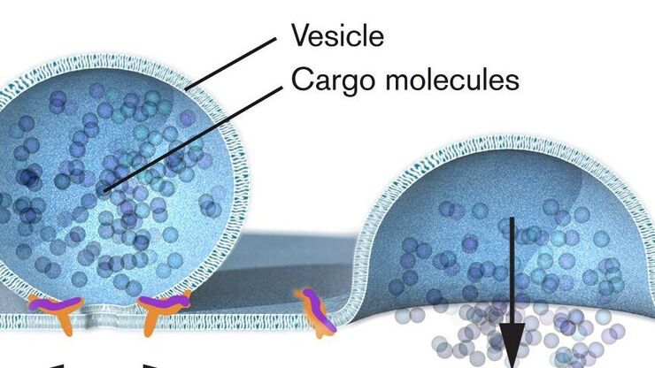
Nobel Prize 2013 in Physiology or Medicine for Discoveries of the Machinery Regulating Vesicle Traffic
On October 7th 2013, The Nobel Assembly at Karolinska Institutet has decided to award The Nobel Prize in Physiology or Medicine 2012 jointly to James E. Rothman, Randy W. Schekman and Thomas C. Südhof…
Loading...

Handbook of Optical Filters for Fluorescence Microscopy
Fluorescence microscopy and other light-based applications require optical filters that have demanding spectral and physical characteristics. Often, these characteristics are application-specific and…
Loading...
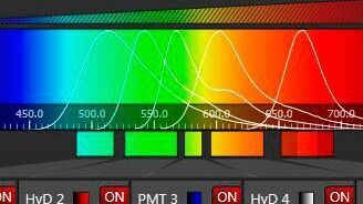
Spectral Detection – How to Define the Spectral Bands that Collect Probe-specific Emission
To specifically collect emission from multiple probes, the light is first separated spatially and then passes through a device that defines a spectral band. Classically, this is a common glass-based…
