
Science Lab
Science Lab
Willkommen auf dem Wissensportal von Leica Microsystems. Hier finden Sie wissenschaftliches Forschungs- und Lehrmaterial rund um das Thema Mikroskopie. Das Portal unterstützt Anfänger, erfahrene Praktiker und Wissenschaftler gleichermaßen bei ihrer täglichen Arbeit und ihren Experimenten. Erkunden Sie interaktive Tutorials und Anwendungshinweise, entdecken Sie die Grundlagen der Mikroskopie ebenso wie High-End-Technologien. Werden Sie Teil der Science Lab Community und teilen Sie Ihr Fachwissen.
Filter articles
Tags
Beitragstyp
Produkte
Loading...

Laser Beam Shaping for Multicolor Multiphoton Microscopy
Multiphoton Microscopy is one of the current hot topics in life science research. The new Leica TCS SP8 DIVE from Leica Microsystems presents a series of beneficial new innovations, including a freely…
Loading...
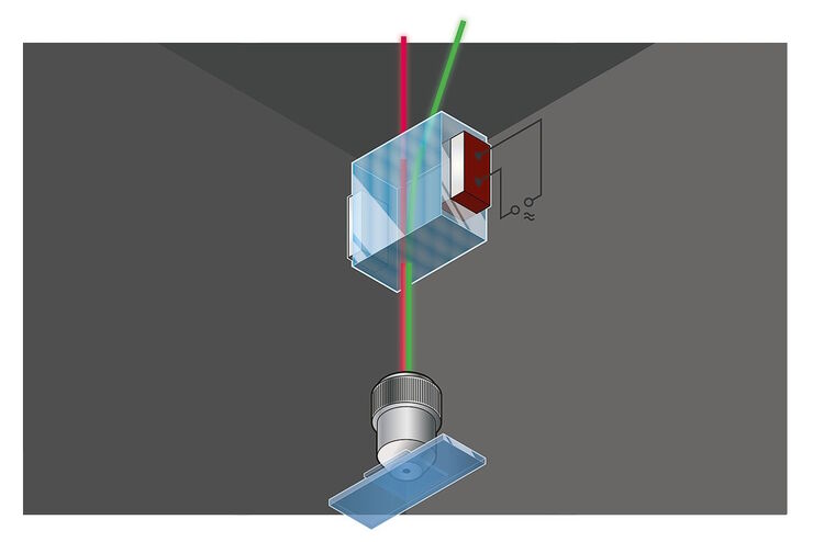
Primary Beam Splitting Devices for Confocal Microscopes
Current fluorescence microscopy employs incident illumination which requires separation of illumination and emission light. The classical device performing this separation is a color-dependent beam…
Loading...

Photoactivatable, Photoconvertible, and Photoswitchable Fluorescent Proteins
Fluorescent proteins (FPs) such as GFP, YFP or DsRed are powerful tools to visualize cellular components in living cells. Nevertheless, there are circumstances when classical FPs reach their limits.…
Loading...

Was macht das Pinhole im konfokalen Mikroskop?
Diese kurze einführende Schrift soll die Bedeutung des Pinholes erläutern und ist für Leser gedacht, die nicht allzu viel Zeit mit der Theorie und den Details der konfokalen Mikroskopie verbringen…
Loading...
 stereo microscope for a task like surgery.")
Rodent and Small-Animal Surgery
Learn how you can perform rodent (mouse, rat, hamster) and small-animal surgery efficiently with a microscope for developmental biology and medical research applications by reading this article.
Loading...
 illuminated with wide-band UV excitation. Note the tissue structure with blue autofluorescence. Right: Same tissue and same immunostaining with FITC label illuminated with epi-illumination using narrow")
Milestones in Incident Light Fluorescence Microscopy
Since the middle of the last century, fluorescence microscopy developed into a bio scientific tool with one of the biggest impacts on our understanding of life. Watching cells and proteins with the…
Loading...

Chronic Inflammation Under the Microscope
In the course of chronic inflammation certain body areas are recurrently inflamed. This goes along with many human diseases. With the help of widefield light microscopy, the underlying processes can…
Loading...

Gene Editing with CRISPR/Cas9 - Breakthrough in Genome Engineering
The CRISPR/Cas9 system is one of several different bacterial systems for defense against viral attacks. It consists of two main components. One is a small piece of RNA which binds to the viral target…
Loading...

Imaging and Analyzing Zebrafish, Medaka, and Xenopus
Discover how to image and analyze zebrafish, medaka, and Xenopus frog model organisms efficiently with a microscope for developmental biology applications from this article.
