
Filter articles
タグ
製品
Loading...

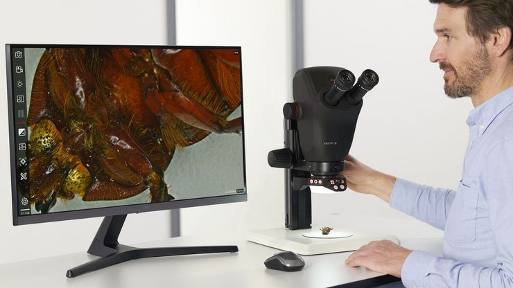
ゼブラフィッシュを用いた研究
スクリーニング、ソーティング、マニピュレーションおよびイメージングを通じて最良の結果を得るためには、細部や構造を観察して、研究の次の段階に向けて正しい判断を下す必要があります。
優れた光学系と高解像度で定評のあるライカの実体顕微鏡と透過照明スタンドは、世界中の研究者から支持されています。
Loading...
")
How to Streamline High-Plex Imaging for 3D Spatial Omics Advances
In this webinar, Dr. Julia Roberti and Dr. Luis Alvarez from Leica Microsystems introduce SpectraPlex, a new functionality integrated into the STELLARIS confocal platform for high-plex 3D spatial…
Loading...

The Guide to Augmented Reality in Microsurgery
In an era of technological advancement, Augmented Reality (AR) is rapidly transforming the medical field. In surgical microscopy, AR can display fluorescence signals as digital overlays in real-time…
Loading...

Improving Zebrafish-Embryo Screening with Fast, High-Contrast Imaging
Discover from this article how screening of transgenic zebrafish embryos is boosted with high-speed, high-contrast imaging using the DM6 B microscope, ensuring accurate targeting for developmental…
Loading...

Transforming Research with Spatial Proteomics Workflows
Spatial Proteomics, Nature Methods 2024 Method of the Year, is driving research advancements in cancer, immunology, and beyond. By combining positional data with high throughput imaging of proteins in…
Loading...
.")
How Fluorescence Guides Sectioning of Resin-embedded EM Samples
Electron microscopes, including transmission electron microscopes (TEM) and scanning electron microscopes (SEM), are widely utilized to gain detailed structural information about biological samples or…
Loading...

Coherent Raman Scattering Microscopy Publication List
CRS (Coherent Raman Scattering) microscopy is an umbrella term for label-free methods that image biological structures by exploiting the characteristic, intrinsic vibrational contrast of their…
Loading...
レーザーマイクロダイセクション
小動物などの解剖を行うとき、顕微鏡の接眼レンズを何時間ものぞくことがあります。 ライカ マイクロシステムズでは、さまざまな顕微鏡と幅広い解剖顕微鏡部品やアクセサリーから選ぶことができるため、ニーズに最適な顕微鏡ソリューションを見つけることができます。
Loading...

Get to Insights Faster and Easier with AI Image Analysis Tools
Discover how Aivia helps scientists streamline image analysis with fast setup, accurate AI detection, and easy batch processing.
