Science Lab

Science Lab

Science Lab

ライカマイクロシステムズのナレッジポータルでは、顕微鏡の基礎から最先端技術まで、幅広い情報を提供しています。初心者から熟練者、研究者、医師の皆様まで、日々の研究や実験に役立つ内容となっております。チュートリアルやアプリケーションノートを活用し、学びながら探究心を刺激してください。さらに、コミュニティに参加することで、知見を共有し、新たな発見へとつなげましょう。お気軽に参加いただき、互いの専門知識を深め合う場としてご活用ください。
Image: Adult rat brain. Neurons (Alexa Fluor488, green), Astrocytes (GFAP, red), Nuclei (DAPI, blue). Image courtesy of Prof. En Xu, Institute of Neurosciences and Department of Neurology of the Second Affiliated Hospital of Guangzhou Medical University, China.

Multicolor Microscopy: The Importance of Multiplexing

The term multiplexing refers to the use of multiple fluorescent dyes to examine various elements within a sample. Multiplexing allows related components and processes to be observed in parallel,…

A New Method for Convenient and Efficient Multicolor Imaging

The technique combining hyperspectral unmixing and phasor analysis was developed to simplify the process of getting images from a sample labeled with multiple fluorophores. This aggregate method…

Considerations for Multiplex Live Cell Imaging

Simultaneous multicolor imaging for successful experiments: Live-cell imaging experiments are key to understand dynamic processes. They allow us to visually record cells in their living state, without…
Virally labeled neurons (red) and astrocytes (green) in a cortical spheroid derived from human induced pluripotent stem cells. THUNDER Model Organism Imagerwith a 2x 0.15 NA objective at 3.4x zoomwas used to produce this 425 μm Z-stack (26 positions), which is presented here as an Extended Depth of Field(EDoF)projection.

Neuroscience Images

Neuroscience commonly uses microscopy to study the nervous system’s function and understand neurodegenerative diseases.
Virally labeled neurons (red) and astrocytes (green) in a cortical spheroid derived from human induced pluripotent stem cells. THUNDER Model Organism Imager with a 2x 0.15 NA objective at 3.4x zoom was used to produce this 425 µm Z-stack (26 positions), which is presented here as an Extended Depth of Field (EDoF) projection.  Images courtesy of Dr. Fikri Birey  from the Dr. Sergiu Pasca laboratory at Stanford University, 3165 Porter Dr., Palo Alto, CA

Download The Guide to Live Cell Imaging

In life science research, live cell imaging is an indispensable tool to visualize cells in a state as in vivo as possible. This E-book reviews a wide range of important considerations to take to…

研究におけるモデル生物

モデル生物とは、特定の生物学的プロセスを研究するために研究者が使用する生物種です。 モデル生物は、人間と似た遺伝的特徴を持ち、遺伝子学、発生生物学、神経科学などの研究分野で一般的に使用されています。 通常、モデル生物は実験環境での維持や繁殖が容易であること、生殖サイクルが短いこと、または、特定の形質や病気を研究するために突然変異体を生成する能力を持つことで選ばれます。
Convalaria

Introduction to Widefield Microscopy

This article gives an introduction to widefield microscopy, one of the most basic and commonly used microscopy techniques. It also shows the basic differences between widefield and confocal…

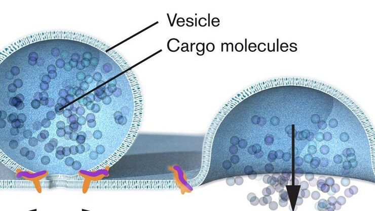
Nobel Prize 2013 in Physiology or Medicine for Discoveries of the Machinery Regulating Vesicle Traffic

On October 7th 2013, The Nobel Assembly at Karolinska Institutet has decided to award The Nobel Prize in Physiology or Medicine 2012 jointly to James E. Rothman, Randy W. Schekman and Thomas C. Südhof…
John B. Gurdon

Nobel Prize 2012 in Physiology or Medicine for Stem Cell Research

The Nobel Prize recognizes two scientists who discovered that mature, specialised cells can be reprogrammed to become immature cells capable of developing into all tissues of the body. Their findings…
Scroll to top