
Filter articles
タグ
製品
Loading...

Observing Complex Cellular Interactions at Multiple Scales
Learn how to observe challenging cellular interactions with easy to deploy object detection and relationship measurements.
Loading...

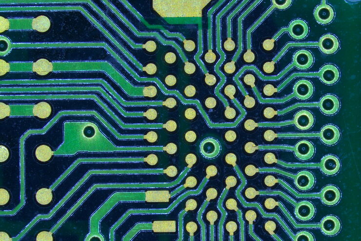
How to Boost your Microelectronic Component Inspection Performance
Do you need to see more when inspecting silicon wafers or MEMS? Would you like to get sharp and detailed sample images which are similar to those from electron microscopes?
Watch this free webinar…
Loading...

How Industrial Applications Benefit from Fluorescence Microscopy
Watch this free webinar to know more about what you can do with fluorescence microscopy for industrial applications. We will cover a wide range of investigations where fluorescence contrast offers new…
Loading...

Intravital Microscopy of Cancer
Join our guest speaker Prof Dr Jacco van Rheenen, as he presents his work on the identity, behavior and fate of cells that drive the initiation and progression of cancer.
Loading...

Accelerating Neuron Image Analysis with Automation
The ability to examine complex neural processes relies on the accurate reconstruction of neuronal networks at scale. Most data extraction methods in neuroscience research are time-consuming and…
Loading...
Tracking Single Cells Using Deep Learning
AI-based solutions continue to gain ground in the field of microscopy. From automated object classification to virtual staining, machine and deep learning technologies are powering scientific…
Loading...
How to Use a Digital Microscope to Streamline Inspection Processes
Watch this webinar for inspiration and expert advice on how to make quality control simpler, quicker, and easier. Learn how to perform comprehensive visual inspection, including comparison,…
Loading...

Designing your Research Study with Multiplexed IF Imaging
Multiplexed tissue analysis is a powerful technique that allows comparisons of cell-type locations and cell-type interactions within a single fixed tissue sample. It is common for researchers to ask…
Loading...

High-resolution 3D Imaging to Investigate Tissue Ageing
Award-winning researcher Dr. Anjali Kusumbe demonstrates age-related changes in vascular microenvironments through single-cell resolution 3D imaging of young and aged organs.
